
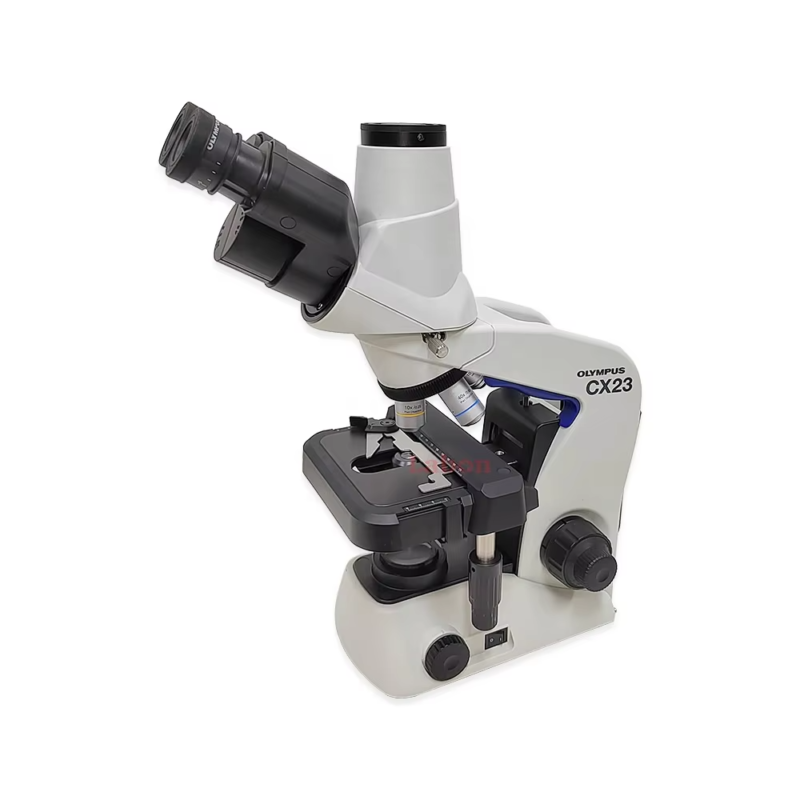
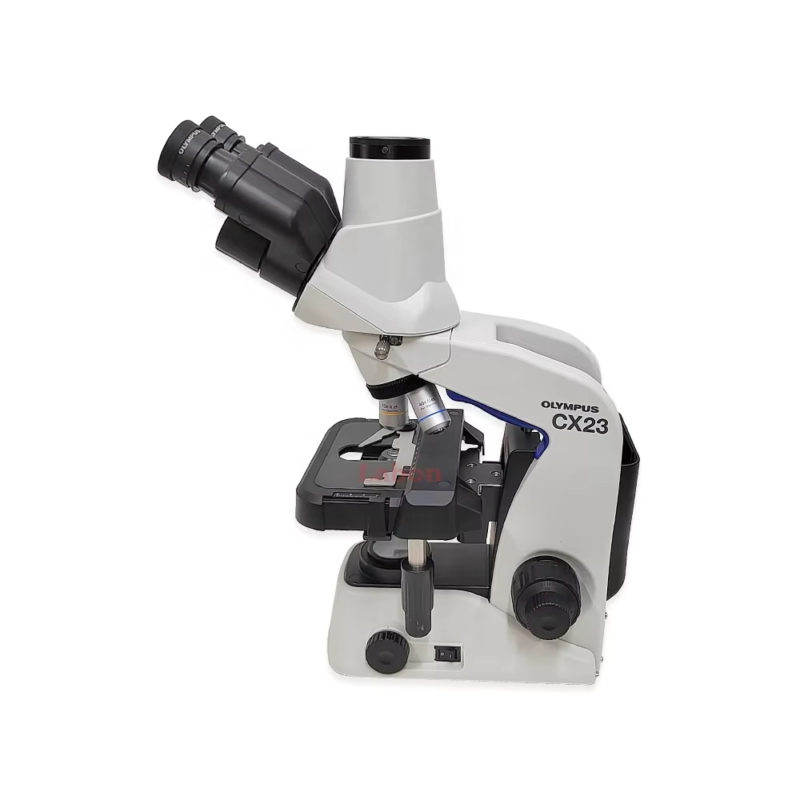
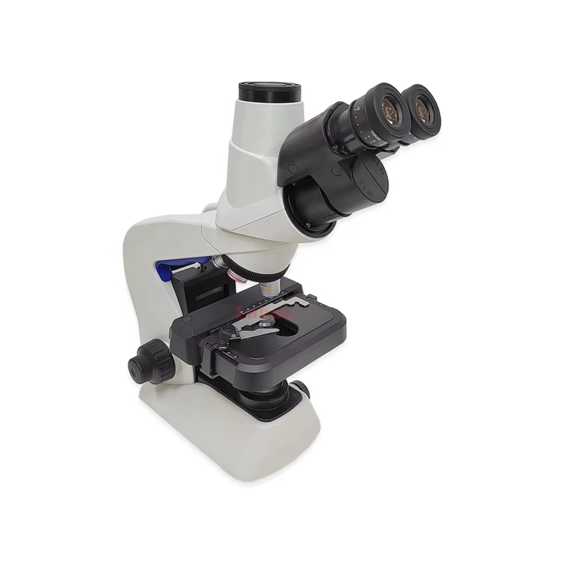
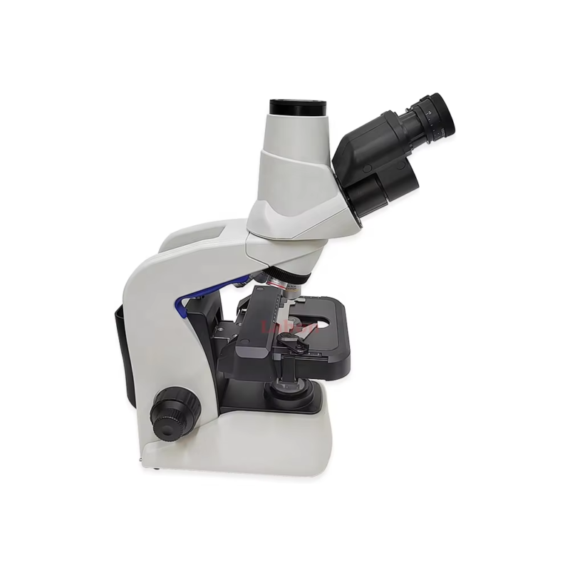
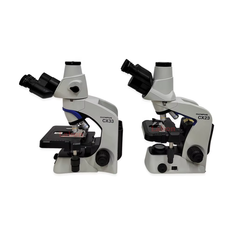
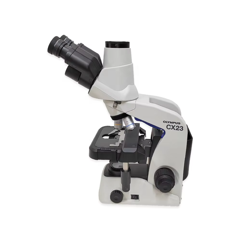
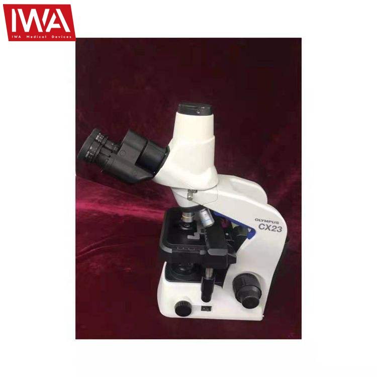
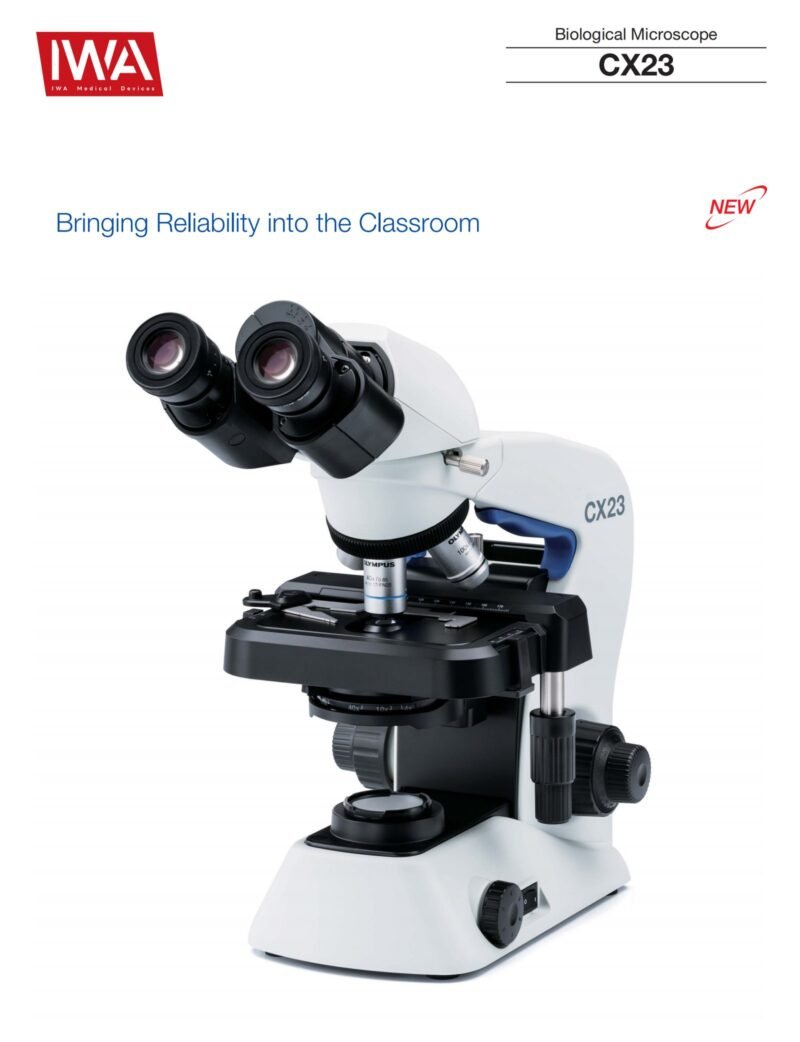
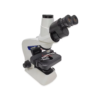
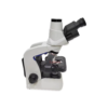
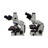
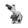
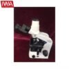
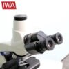
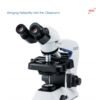
Olympus CX23 trinocular biological microscope — same plan achromat optics as the binocular model, with a camera port for documenting cytology and referral images.
Product Overview
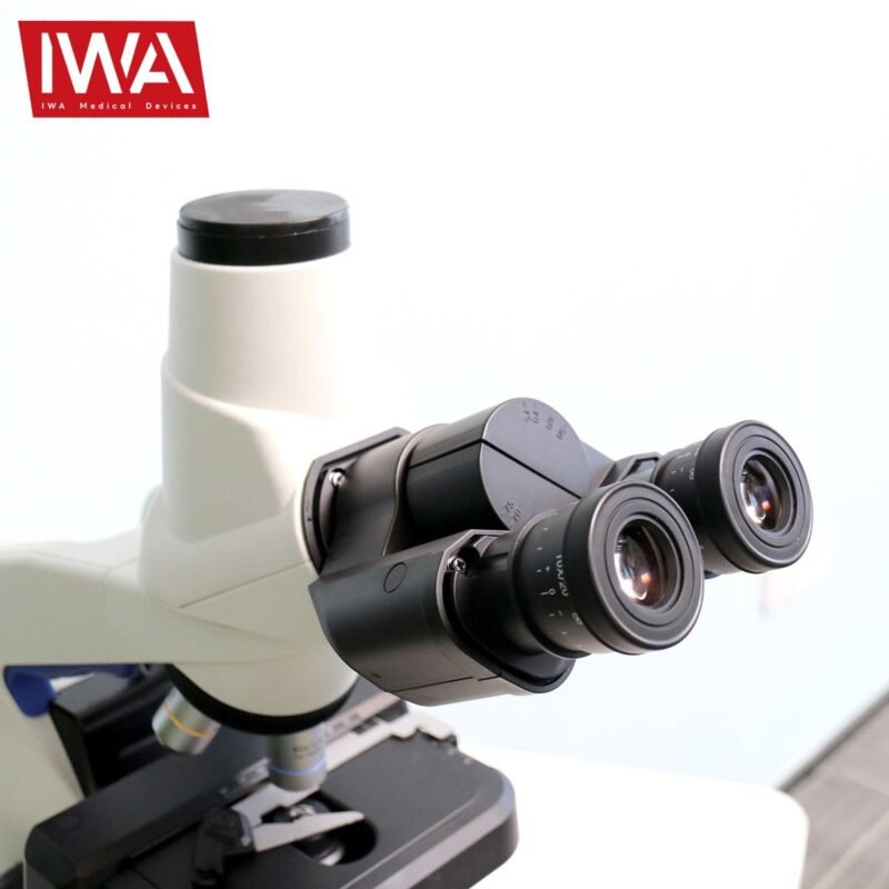
The trinocular CX23 carries the same infinity optical system, plan achromat objectives and FN20 eyepieces as the binocular model, with a third tube for a camera. The light path selector sends 100% to the eyepieces or 100% to the camera, so image capture does not compromise what the operator sees.
Supplied as CX23RTFS2, which includes the CX23-FS field diaphragm as standard. Choose this version if the practice records cytology findings in the patient file, sends images for referral opinion, or teaches from the microscope.
Key Features
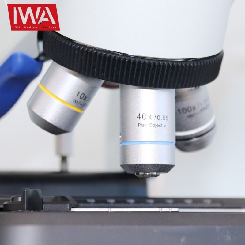
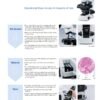
- Plan achromat objectives as standard (4×/10×/40×/100× oil) — image stays flat across the full field, no centre-only focus
- Field number 20 eyepieces — wider field than FN18 optics, fewer passes to scan a smear
- Infinity optical system on an aluminium die-cast frame with protective plastic covering
- Abbe condenser NA 1.25 with oil immersion and built-in aperture iris for contrast control
- LED illumination at 0.5 W, 20,000-hour rated life and daylight-balanced colour — holds true colour on H&E and Diff-Quik stains
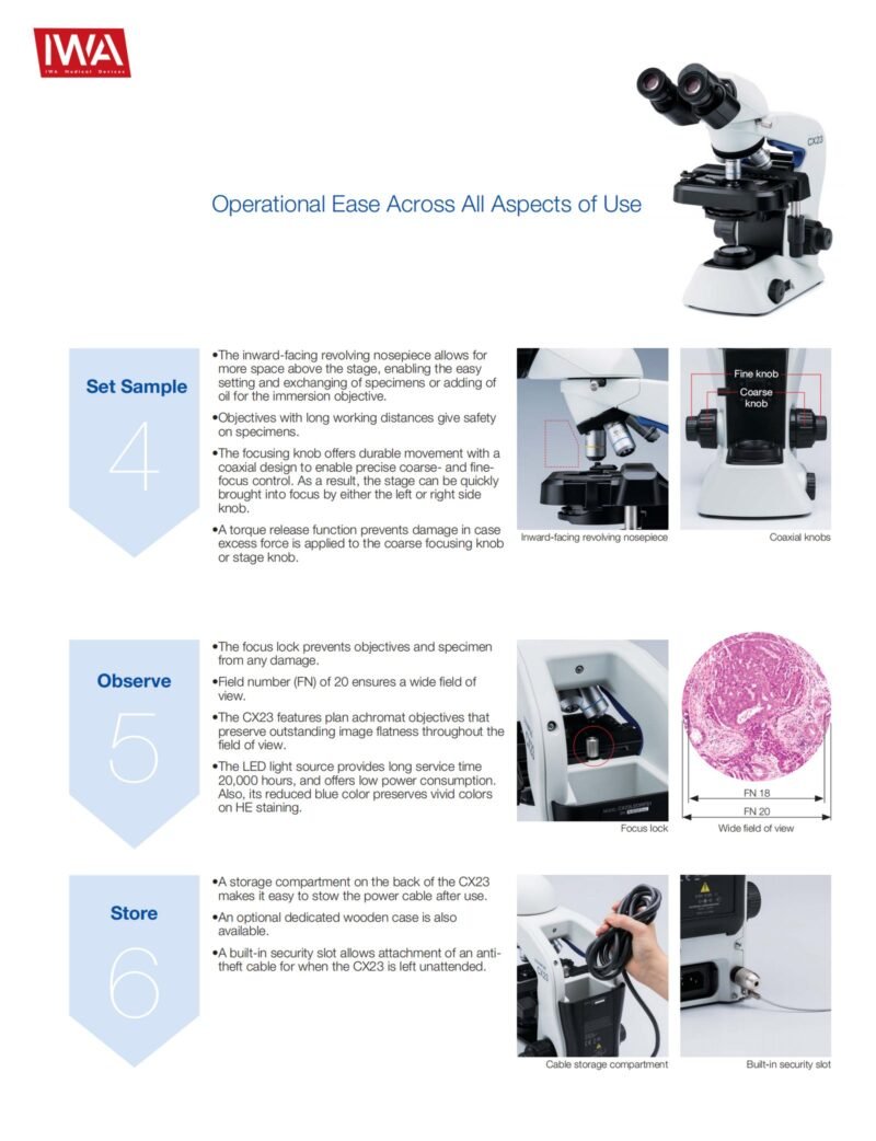
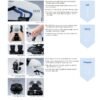
- Coaxial coarse / fine focus, 15 mm coarse stroke, 2.5 µm minimum fine graduation, with focus lock and torque adjustment
- Torque release on the coarse and stage knobs limits damage if excess force is applied
- Binocular tube 30° inclined, interpupillary distance 48–75 mm, left and right diopter adjustment
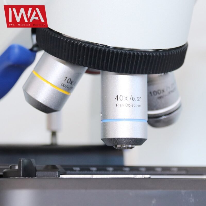
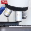
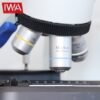
- Inward-tilting quadruple nosepiece — clears working space above the stage for slide changes and oiling
- Rackless mechanical stage, 76 × 30 mm travel, with specimen holder and position scale
- Light path selector — 100/0 or 0/100 between eyepieces and camera port
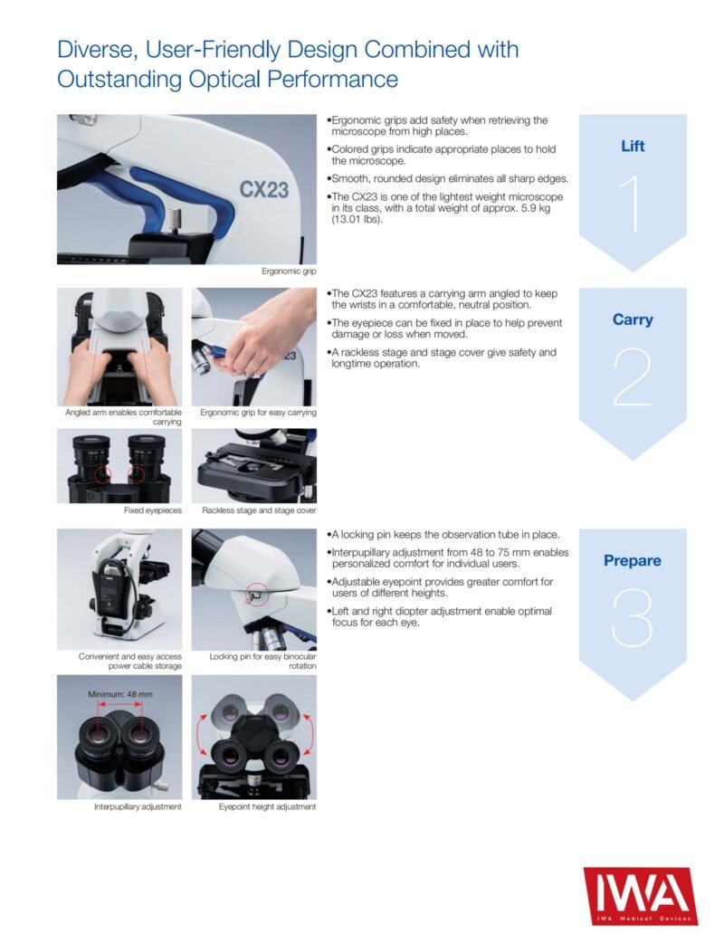
- 6.5 kg with ergonomic grips and cable storage — movable between consulting rooms
Technical Specifications
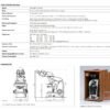
| Brand | Olympus (Japan) |
| Model | CX23RTFS2 (trinocular) |
| Optical system | Infinity optical system |
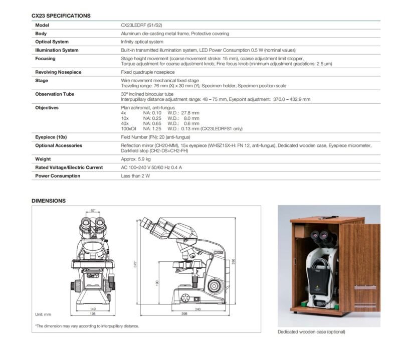
| Observation tube | Trinocular, anti-fungal, 30° inclined; interpupillary distance 48–75 mm |
| Light path selector | Eyepiece / camera port = 100/0 or 0/100 |
| Eyepieces | 10× anti-fungal, field number (FN) 20; 15× (FN 12) available as an option; eyepoint 370.0–432.9 mm |
| Objectives | Plan achromat, anti-fungal — 4× (NA 0.1, WD 27.8 mm), 10× (NA 0.25, WD 8.0 mm), 40× (NA 0.65, WD 0.6 mm), 100× oil (NA 1.25, WD 0.13 mm) |
| Nosepiece | Fixed quadruple nosepiece, inward tilt |
| Stage | Wire-movement mechanical fixed stage, 174 × 89 mm; travel 76 × 30 mm (X × Y); specimen holder and position scale |
| Condenser | Abbe condenser NA 1.25 with oil immersion; built-in aperture iris diaphragm |
| Focusing | Stage height movement; coarse stroke 15 mm with limit stopper and torque adjustment; fine focus minimum graduation 2.5 µm |
| Illumination | Built-in transmitted LED, 0.5 W nominal, 20,000-hour lifetime |
| Power | AC 100–240 V, 50/60 Hz, 0.4 A; total consumption under 2 W |
| Dimensions (W × D × H) | 198 × 398 × 430 mm |
| Weight | Approx. 6.5 kg |
| Optional accessories | Reflection mirror (CH20-MM), eyepiece micrometer, dust cover, darkfield filter (CH2-DS), dedicated wooden case (CH20-WB2) |
Clinical Applications
- Routine veterinary cytology (FNA, ear, urine sediment)
- Hematology blood smear examination
- Urinalysis sediment examination
- Fecal flotation and direct smear parasitology
- Veterinary teaching laboratories
Package Contents
- Olympus CX23 trinocular microscope body (CX23RTFS2)
- 10× FN20 eyepieces × 2
- Plan achromat objectives 4× / 10× / 40×
- CX23-FS field diaphragm — included in the CX23RTFS2 standard set
- 100× oil immersion objective — optional on this set
- AC power cable
- User manual
Why Choose CX23
- Plan achromat optics as standard — flat field across FN20 means fewer passes per smear and less refocusing at the edges
- Focus lock and torque release protect objectives and slides in a shared-use practice laboratory
- LED at 0.5 W and 20,000 hours — no bulb stock to hold, no colour shift as the lamp ages
- Camera port with 0/100 switching — capture a referral image without the operator losing the field
- Under 2 W total draw and 198 × 398 mm bench footprint — no dedicated laboratory bench required
Warranty & Support
FAQ
Which veterinary cytology / histology slides does this microscope read? Routine veterinary cytology smears (FNA, ear cytology, urine sediment), histopathology slides, fecal flotation / direct smear, urinalysis sediment, and hematology (Diff-Quik / Wright-Giemsa stained) workflow.
How long does the LED last? The LED is rated at 20,000 hours and draws 0.5 W. At eight hours a day that is roughly seven years of use. Replacement is a service operation — contact us rather than opening the base.
Can objectives from another microscope be fitted? The CX23 uses an infinity optical system, so objectives computed for a finite tube length will not focus correctly on it. Use objectives specified for this infinity series; the 20× plan achromat (NA 0.4, WD 2.5 mm) is available as an option.
Which camera fits the third tube? The camera port takes an Olympus camera adapter matched to the sensor size; the adapter is not included. Tell us the camera or tablet system the practice uses and we will confirm the correct adapter before ordering.
Do I lose brightness in the eyepieces when the camera is fitted? No. The selector is 100/0 or 0/100, not a beam splitter — light goes either to the eyepieces or to the camera, at full intensity.
Does it need a dedicated bench or special power? No. It runs on AC 100–240 V, 50/60 Hz, draws under 2 W, occupies 198 × 398 mm of bench and weighs 6.5 kg. The power cable stows in a compartment at the back.

Reviews
There are no reviews yet.