
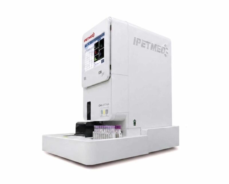
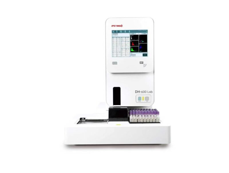
Automated veterinary hematology analyzer delivering 5-part leukocyte differential, reticulocyte analysis and nucleated-RBC detection by tri-angle laser flow cytometry with nucleic-acid fluorescence staining.
Hero Media
Product Overview
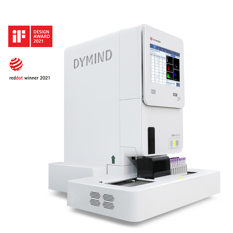
IWA-DH600LAB is a fully automated veterinary blood cell analyzer built for diagnostic and research laboratories. It combines three-angle laser flow cytometry with nucleic-acid fluorescence staining to deliver an accurate 5-part white blood cell differential, reticulocyte typing, immature granulocyte and nucleated-RBC detection, and abnormal-cell flagging. The system pairs a benchtop main unit with a 60-tube automatic sampler, an external air source, on-board software and PC software, and dedicated reagents — running CBC, RET and combined modes from a single platform.
Key Features
- 5-part WBC differential with reticulocyte typing and nucleated-RBC (NRBC) detection — no off-instrument staining required
- 35 reportable parameters across five modes (CBC, RET, CBC+DIFF, CBC+RET, CBC+DIFF+RET), plus 29 research parameters
- Tri-angle laser flow cytometry with nucleic-acid fluorescence staining for DIFF and RET; impedance for WBC/Baso/RBC; dual impedance-plus-optical PLT; cyanide-free colorimetric HGB
- Throughput up to 100 samples/hour (CBC); full CBC+DIFF+RET workup at 66 samples/hour
- Low sample volume — CBC from ≤20 µL venous or micro whole blood
- 60-tube continuous auto-sampler with STAT interrupt, closed-tube piercing and automatic micro-blood sampling
- Dedicated low-WBC and low-PLT modes raise counted particle numbers for reliable results at low concentrations
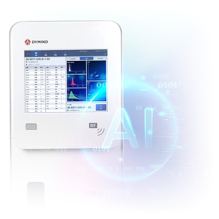
- On-board AI analysis flags abnormal cells, provides disease-risk alerts and suggests diagnostic direction
Visual Evidence
Technical Specifications
| Model | DH-600Lab |
| Methodology — DIFF / RET | Three-angle laser flow cytometry with nucleic-acid fluorescence staining (3D analysis) |
| Methodology — WBC / Baso / RBC | Impedance |
| Methodology — PLT | Dual: impedance and optical fluorescence |
| Methodology — HGB | Cyanide-free colorimetric |
| Reportable parameters | 35 |
| Research parameters | 29 |
| Analysis modes | CBC, RET, CBC+DIFF, CBC+RET, CBC+DIFF+RET |
| Detection channels | WBC differential, WBC/Baso/HGB, reticulocyte, RBC/PLT impedance |
| Graphics | 3 histograms, 2 three-dimensional scattergrams, 8 two-dimensional scattergrams |
| Sample types | Venous whole blood, micro whole blood, pre-dilute |
| Auto-sampler capacity | 60 tubes, continuous uninterrupted loading; STAT interrupt |
| Sampling modes | Full auto, single closed-tube, single open-cap; auto micro-blood piercing |
| Display | 12.1″ colour LCD touchscreen; external PC operation supported |
| Data storage | ≥150,000 sample records (with scattergrams, histograms, patient info) |
| QC | L-J, X-B and multi-rule; automatic QC charting |
| Calibration | Calibrator, fresh blood, manual |
| Connectivity | 4 × USB, 1 × LAN; bidirectional HL7 LIS |
| Barcode | Built-in 360° auto sample barcode scanner; external scanner supported |
| Reference ranges | 8 preset population groups by sex/age; custom groups supported |
| Power | AC 100–240 V, 50/60 Hz |
| Operating environment | 15–32 °C, RH 30–85%, 70–106 kPa |
| Validated species | 20+ research and companion species (rat, mouse, guinea pig, rabbit, canine, bovine, equine, ovine, porcine, non-human primate, others) |
Throughput by mode (venous / micro whole blood, samples per hour)
| Mode | Venous | Micro |
|---|---|---|
| CBC | 100 | 90 |
| RET | 66 | 55 |
| CBC+DIFF | 90 | 70 |
| CBC+RET | 66 | 55 |
| CBC+DIFF+RET | 66 | 55 |
Sample volume by mode (venous / micro whole blood)
| Mode | Venous | Micro |
|---|---|---|
| CBC | ≤20 µL | ≤20 µL |
| RET | ≤35 µL | ≤26 µL |
| CBC+DIFF | ≤30 µL | ≤21 µL |
| CBC+RET | ≤35 µL | ≤26 µL |
| CBC+DIFF+RET | ≤35 µL | ≤26 µL |
Key performance indicators
| Parameter | Background (blank) | Repeatability CV | Linearity | Carryover |
|---|---|---|---|---|
| WBC | ≤0.1 ×10⁹/L | ≤2.5% | (0–500) ×10⁹/L | ≤1.0% |
| RBC | ≤0.02 ×10¹²/L | ≤1.5% | (0–8.60) ×10¹²/L | ≤1.0% |
| HGB | ≤1 g/L | ≤1.0% | (0–260) g/L | ≤1.0% |
| PLT | ≤5 ×10⁹/L | ≤4.0% | (0–5000) ×10⁹/L | ≤1.0% |
| HCT | ≤0.5% | ≤1.5% | (0–75.0)% | ≤1.0% |
| MCV | — | ≤1.0% | — | — |
| RET% | — | ≤15.0% | (0–30.00)% | — |
Clinical Applications
- Companion-animal complete blood counts — canine and feline
- Large-animal and production-species hematology — equine, bovine, ovine, porcine
- Laboratory-animal research — rat, mouse, guinea pig, rabbit, non-human primate
- Reticulocyte analysis and regenerative-anemia assessment with maturity indices
- Anemia, inflammation and infection screening using NRBC, immature granulocyte (IG) and IPF flags
- Suspect-cell flagging — blasts, immature granulocytes, abnormal/atypical lymphocytes, platelet aggregation, RBC agglutination
Workflow / Installation Notes
- Sample types: venous whole blood, micro whole blood and pre-dilute; supports closed micro-blood collection tubes from at least three mainstream brands
- Automatic clog detection and clearing; visualised status view of sensors, temperature, pressure, voltage and current
- Reagent management displays name, lot, expiry and remaining volume; reagent kit comprises two lytic reagents, two stains and two diluents
- RET channel can be disabled to reduce running cost when reticulocyte analysis is not required
- Expandable to a dual-unit hematology cascade, a hematology + immunoassay cascade, or a cabinet track line
- Operating environment 15–32 °C, RH 30–85%, 70–106 kPa; power AC 100–240 V (50/60 Hz); external air source
Package Contents
- DH-600Lab main analyzer unit
- Automatic sampling device (60-tube)
- External air source
- On-board host software and PC software
- Reagent kit — two lytic reagents, two stains, two diluents
- User manual
Downloads / Documentation
Brochure, specification sheet and user manual available on request.
Why Choose IWA-DH600LAB
- Workflow efficiency — 60-tube continuous loading with STAT interrupt, ≥150,000-record storage and bidirectional HL7 LIS keep a busy laboratory moving
- Clinical accuracy — tri-angle laser flow cytometry with fluorescence staining, dedicated low-WBC/low-PLT modes and cyanide-free HGB chemistry
- Practice economics — separable, disable-able RET channel and on-screen reagent management optimise cost per result
Warranty & Support
Standard manufacturer warranty [TBC]. Installation, application training, consumables supply and technical support provided through the distributor network.
FAQ
How many species can the analyzer report? The platform is validated across more than 20 research and companion-animal species, including rat, mouse, guinea pig, rabbit, canine, bovine, equine, ovine, porcine and non-human primate.
Does reticulocyte analysis need manual staining? No. Reticulocyte detection and typing are fully automated on the dedicated RET channel with maturity indices — no off-instrument staining is required.
What is the smallest sample volume? A CBC runs from ≤20 µL of venous or micro whole blood; a full CBC+DIFF+RET workup uses ≤35 µL (venous) or ≤26 µL (micro).
Can it integrate with a laboratory information system? Yes. The unit provides bidirectional HL7 LIS communication over LAN, plus four USB ports and built-in 360° barcode scanning.
Can running cost be reduced when reticulocytes are not needed? Yes. The RET channel can be disabled to save reagent and running cost, and reagent management tracks lot, expiry and remaining volume on screen.
Reviews
There are no reviews yet.