
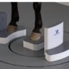

Standing cone-beam CT for the horse — a floor-level ring that scans in as little as 15 s, without general anaesthesia.
Product Overview
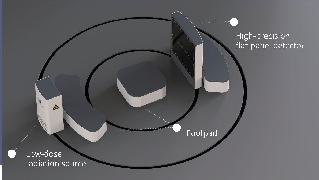
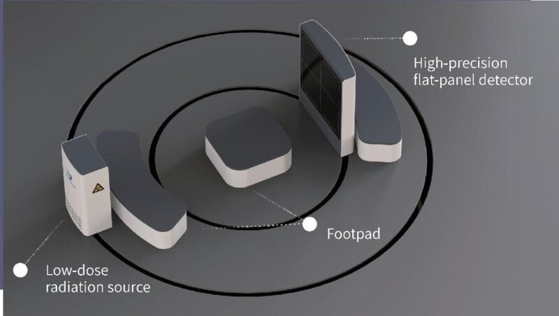
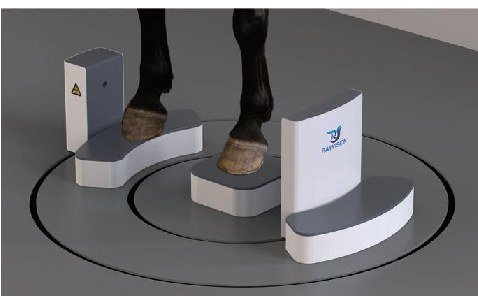
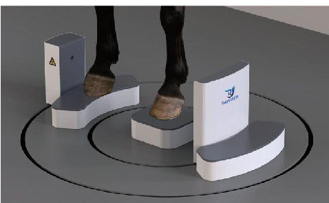
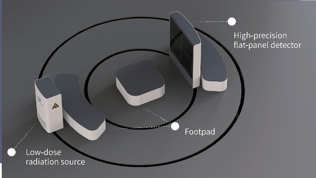
Eq200 is a standing equine cone-beam CT system from Rayvision Medical. A low-dose X-ray source and a high-precision flat-panel detector rotate on a floor-level ring around a central footpad, so the horse is imaged standing in a natural position.
Standing acquisition avoids general anaesthesia and recumbent positioning. Scans complete in as little as 15 s to limit motion artefact, and the field of view is 200 mm (XY) × 165 mm (Z) with slice thickness down to 0.05 mm.
Key Features
- Designed for large equines — standing imaging in a natural position, without general anaesthesia or recumbent positioning.
- Fast scanning — scan time from 15 s (range 15 – 60 s) reduces motion artefact.
- Low radiation dose — low-dose source minimises exposure risk for operators.
- High-resolution volume — 0.05 – 0.24 mm slices, 600 reconstructed slices, 140 µm detector pixel.
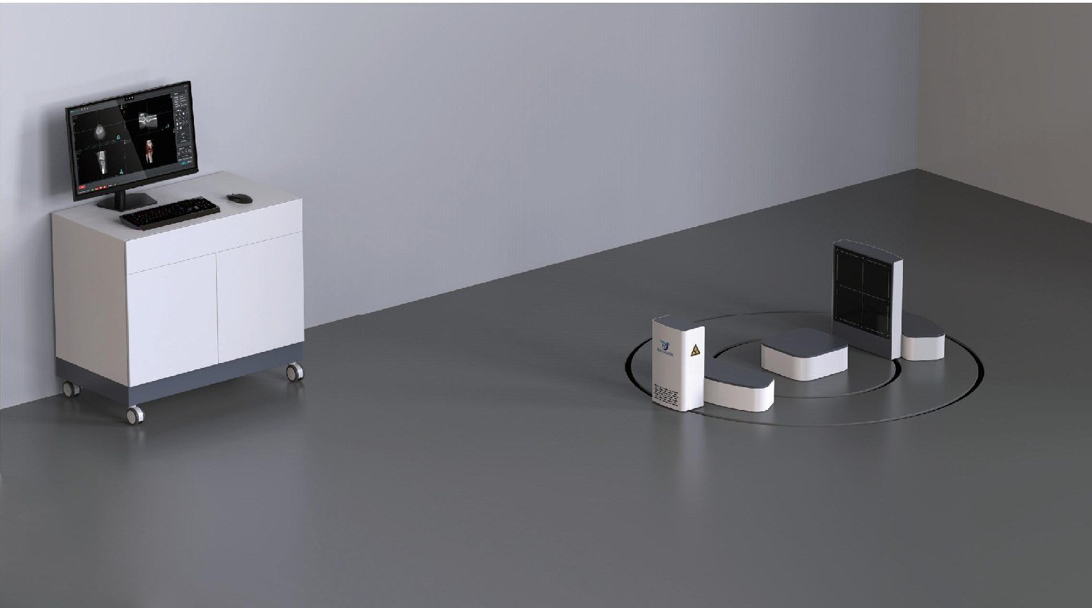
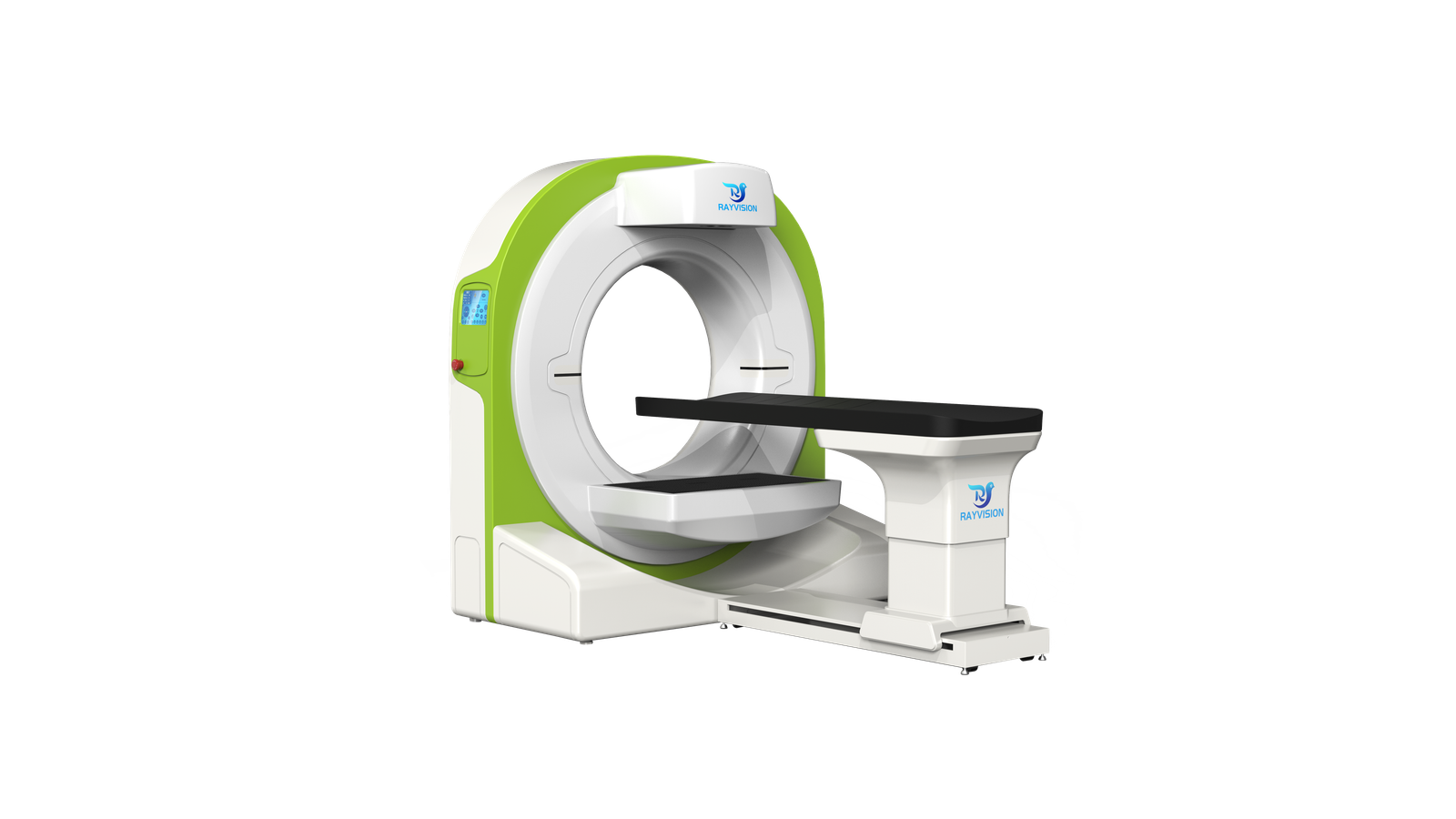
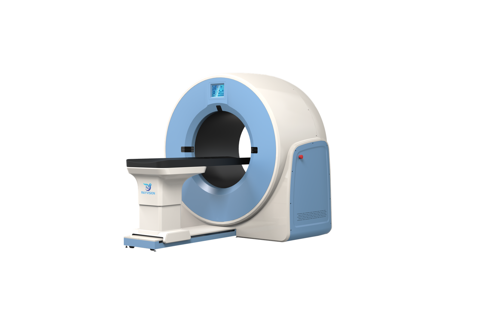
- Floor-level ring gantry — source, footpad and 30 × 30 cm flat-panel detector on one scanning ring.
- 1 kW power requirement — 220 VAC ±10 % supply.
Product Gallery
Technical Specifications — X-ray Source
| Tube voltage | 30 – 80 kV |
| Tube current | 0.5 – 2 mA |
Technical Specifications — Power Supply
| Rated power | 1 kW |
| Input power | 220 VAC ±10 % |
Technical Specifications — Imaging Parameters
| Reconstructed slices | 600 |
| Slice thickness | 0.05 – 0.24 mm |
| FOV (XY) | 200 mm |
| FOV (Z) | 165 mm |
| Scan time | 15 – 60 s |
| Detector pixel | 140 µm |
| Maximum acquisition speed | 30 fps |
| Detector active area | 30 × 30 cm |
Technical Specifications — Ergonomics
| CT gantry weight | 400 kg |
Clinical Applications
- Standing equine CT — the limb to be examined is positioned on the central footpad between the X-ray source and the detector.
Site Planning and Installation
Eq200 is a floor-level installation.
Power and Environment
| Supply voltage | 220 VAC ±10 % |
| Supply internal resistance | ≤ 0.5 Ω |
| Frequency | 50 Hz / 60 Hz |
| Power capacity | 1 kW |
| Operating temperature | 10 °C – 35 °C |
| Relative humidity | 30 % – 75 %, non-condensing |
| Atmospheric pressure | 700 – 1060 hPa |
| Room shielding and dimensions | [TBC] |
Workflow Notes
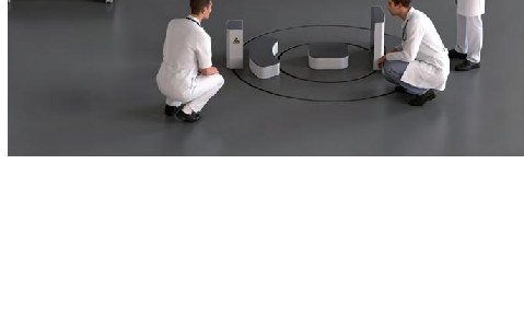
- The horse stands with the limb of interest on the central footpad; sedation is at the attending veterinarian’s discretion.
- The source and detector rotate around the footpad; scans take 15 – 60 s.
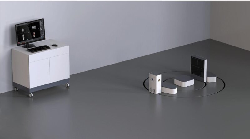
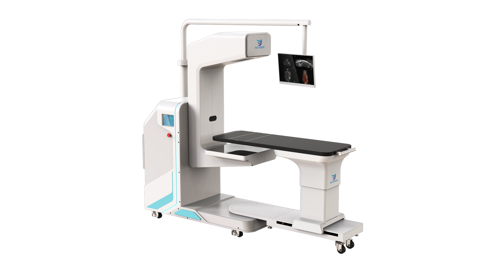
- Images are reviewed at the operator workstation beside the scanning ring.
Package Contents
- 1 × Low-dose X-ray source unit
- 1 × High-precision flat-panel detector unit
- 1 × Footpad
- 1 × Operator workstation
Downloads
Product brochures and technical documentation are available on request — contact IWA.
Why Choose Eq200
- Workflow efficiency — Standing acquisition removes general anaesthesia induction, recumbent positioning and recovery from the imaging workflow.
- Clinical accuracy — Scans from 15 s limit motion artefact; 0.05 mm slices and a 140 µm detector pixel resolve fine osseous detail.
- Practice economics — A 1 kW power requirement and a low-dose source keep facility and operating demands modest.
Warranty and Support
1-year whole-system warranty. Extended warranty is available for purchase — contact IWA for terms and service coverage in your region.
Installation is carried out against the manufacturer’s installation manual: the main unit and patient table are connected, the console and workstation are cabled, and scanning, reconstruction, fluoroscopy and DR acquisition are verified before handover.
FAQ
Q: Does the horse need general anaesthesia?
No. Eq200 images the horse standing in a natural position. Any sedation is at the attending veterinarian’s discretion.
Q: Which anatomy can be scanned?
The limb is positioned on the central footpad between the source and detector, with a 200 mm (XY) × 165 mm (Z) field of view. Contact IWA for the confirmed list of supported regions.
Q: What site preparation is required?
The system needs a 220 VAC ±10 %, 1 kW supply. Shielding, room size and floor preparation for the 400 kg gantry will follow the manufacturer’s site-planning guide — contact IWA. Both 50 Hz and 60 Hz supplies are supported.
Q: What warranty applies?
1-year whole-system warranty, with extended warranty available for purchase.
Reviews
There are no reviews yet.