


Dual-stand digital X-ray system for large animals — 80 kW generator, 1000 mA tube current, and auto-tracking chest stand for equine, bovine, and zoo patients.
Product Overview
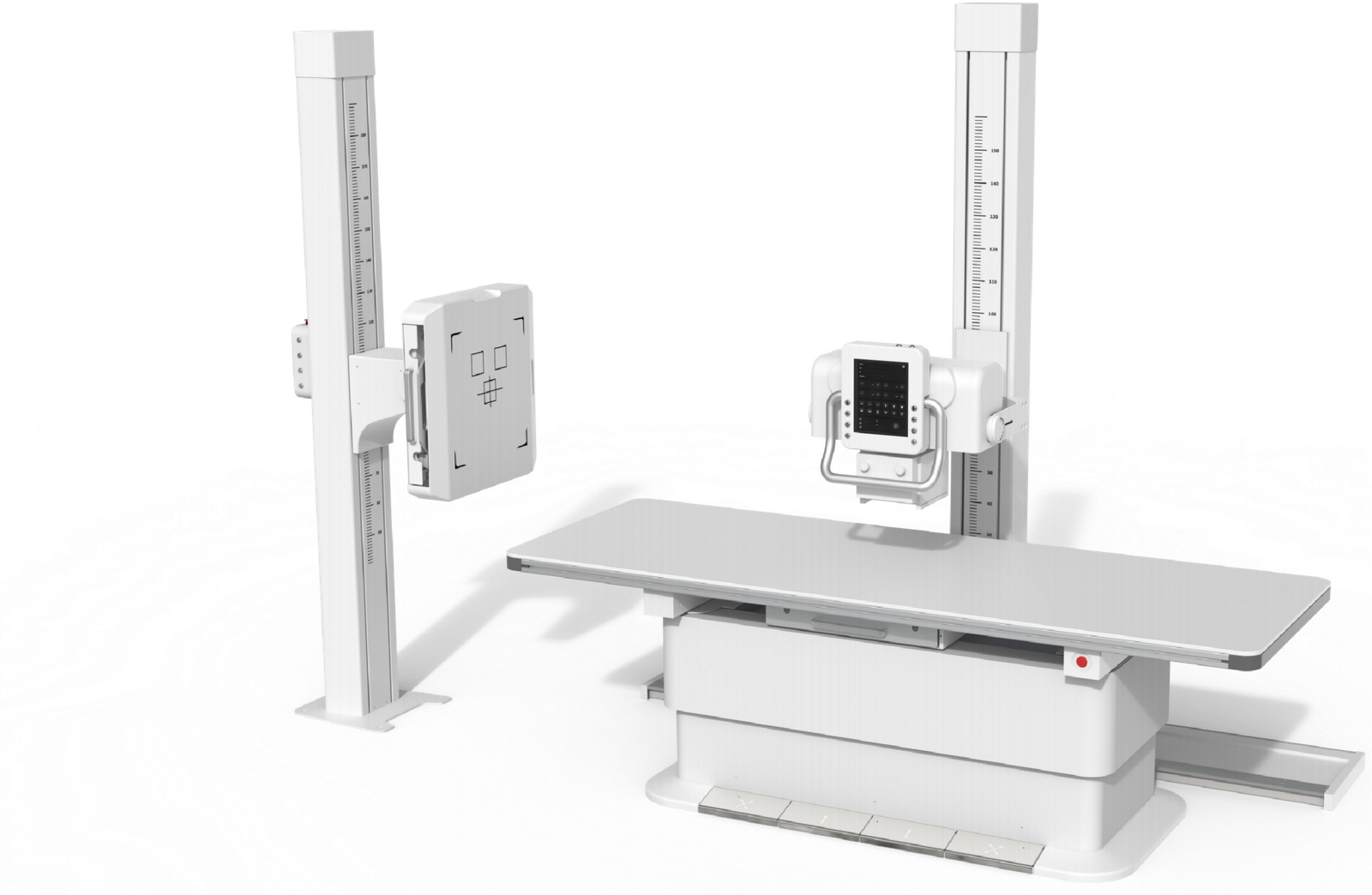
IWA-DR07 iVision-1000X Dual-Stand Digital X-ray System is a dedicated large-animal radiography platform built around an 80 kW high-frequency generator, a high-output 1000 mA X-ray tube, and a 17×17″ CsI flat-panel detector. The dual-track chest stand auto-follows the tube head for fast lateral and AP positioning of horses, cattle, large dogs, and zoo patients.
iVision-1000X is configured for large-animal hospitals, equine and bovine practices, university teaching hospitals, zoos, and wildlife rescue centres that need a fixed-room radiography solution scaled for body masses well beyond small-animal DR systems.
Key Features
- 80 kW high-frequency generator: 40–150 kV, 10–1000 mA, 1–10000 ms exposure, 0.4–1000 mAs — full output range for equine and bovine work.
- Three-phase 380 VAC supply, 135 kVA capacity, 300 kHz inverter for high power density and short exposures.
- High-heat-capacity X-ray tube: 1.0 / 0.6 mm dual focus, rhenium-tungsten on molybdenum anode, 300 kHU anode heat capacity, 1250 kHU component heat capacity.
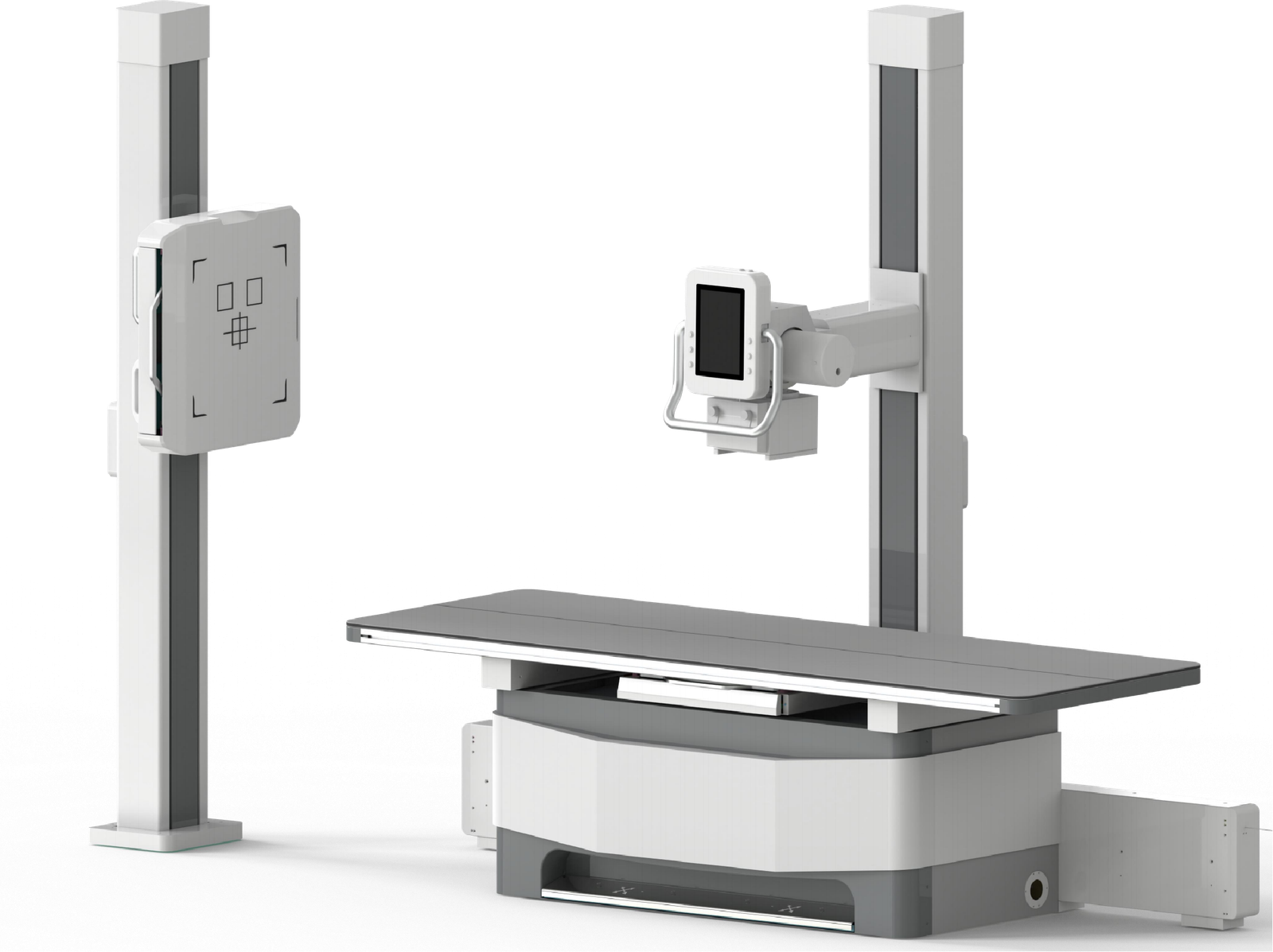
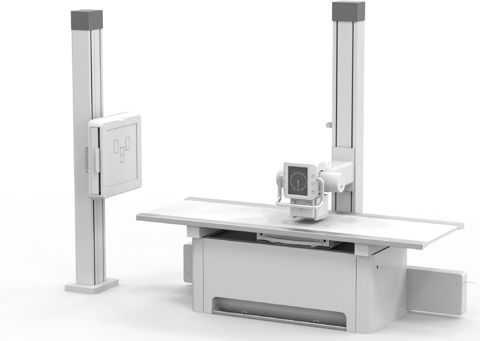
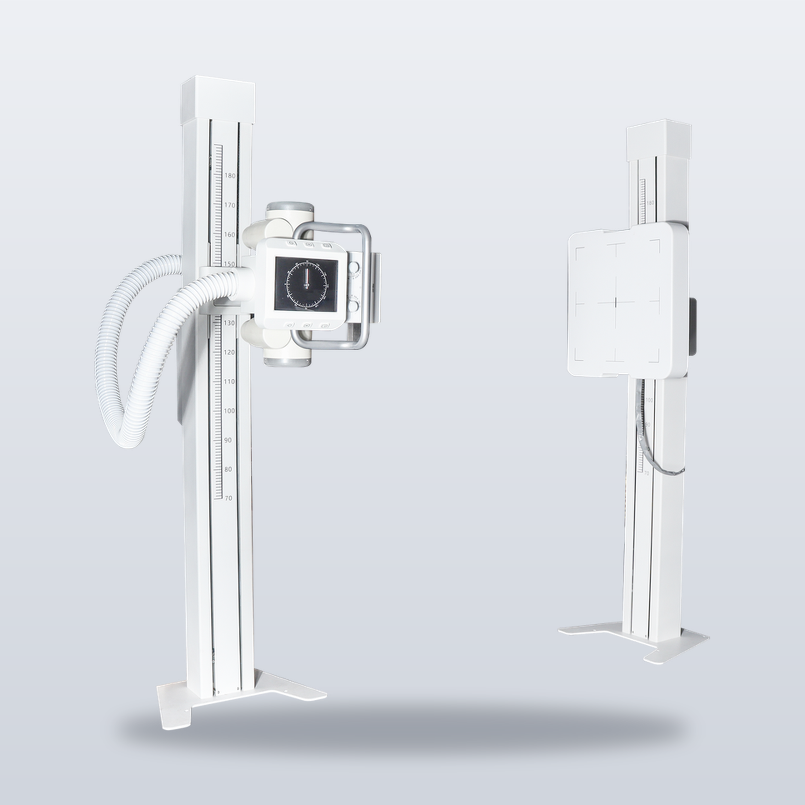
- Dual-track auto-tracking chest stand: vertical travel 1500 mm, grid 10:1 ratio at 180 cm focal length, 103 LPI density.
- Tube stand with full motion: ±180° tube rotation, 1300 mm vertical column travel, 2500 mm horizontal travel along the track.
- 17×17″ CsI flat-panel detector: 3072×3072 matrix, 139 µm pitch, 16-bit A/D, ≥ 3.6 LP/mm spatial resolution.
- iVision Imaging workstation with DICOM 3.0, advanced post-processing, and full veterinary measurement toolkit.
- ABS automatic brightness control during fluoroscopy (when supplied with a dynamic detector).
- Cloud reporting and remote diagnostic workstation support.
- AI-assisted report drafts — the AI imaging assistant drafts, the veterinarian reviews and signs off.
Configuration Options
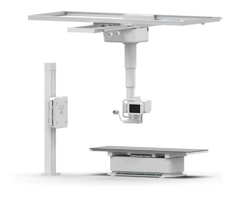
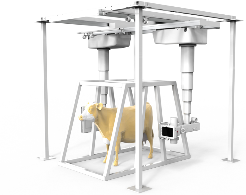
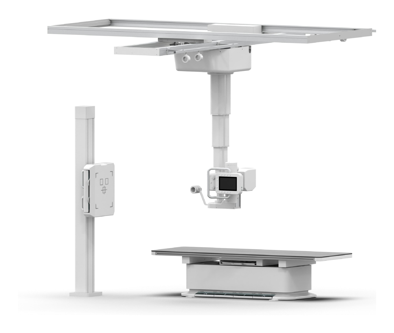
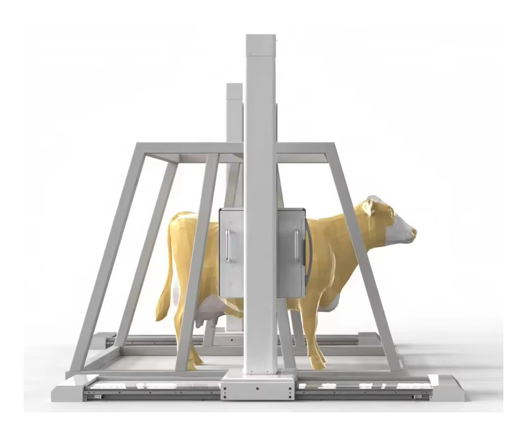
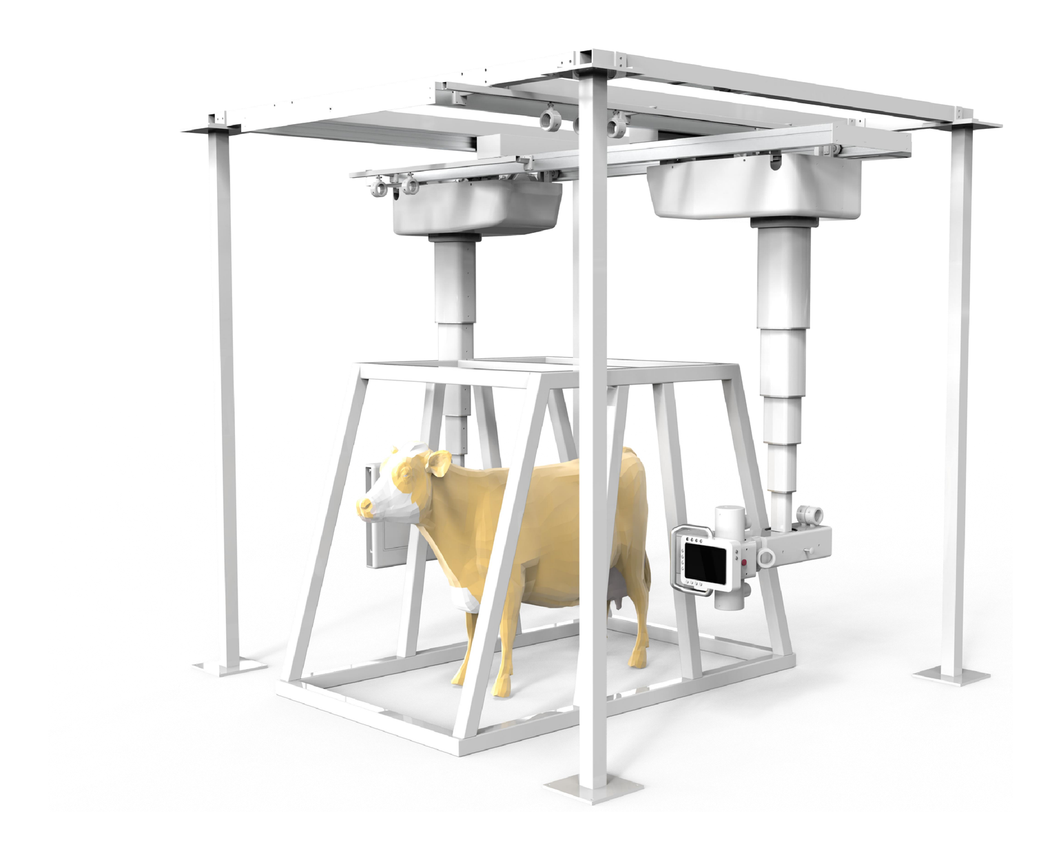
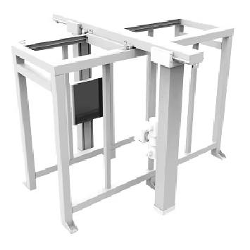
iVision-1000 is supplied in several gantry configurations; the final layout is confirmed in the project proposal for each site:
Product Gallery
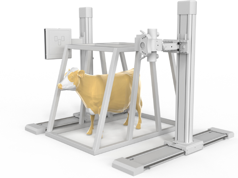
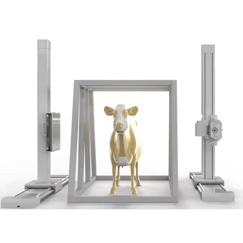
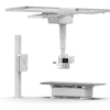
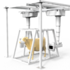
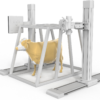
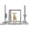
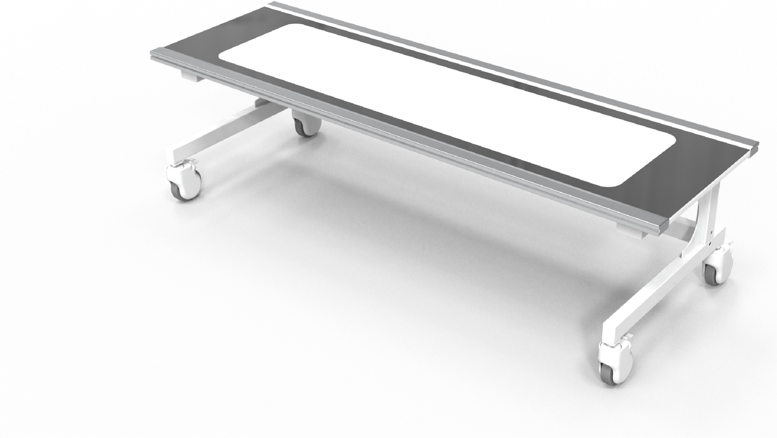
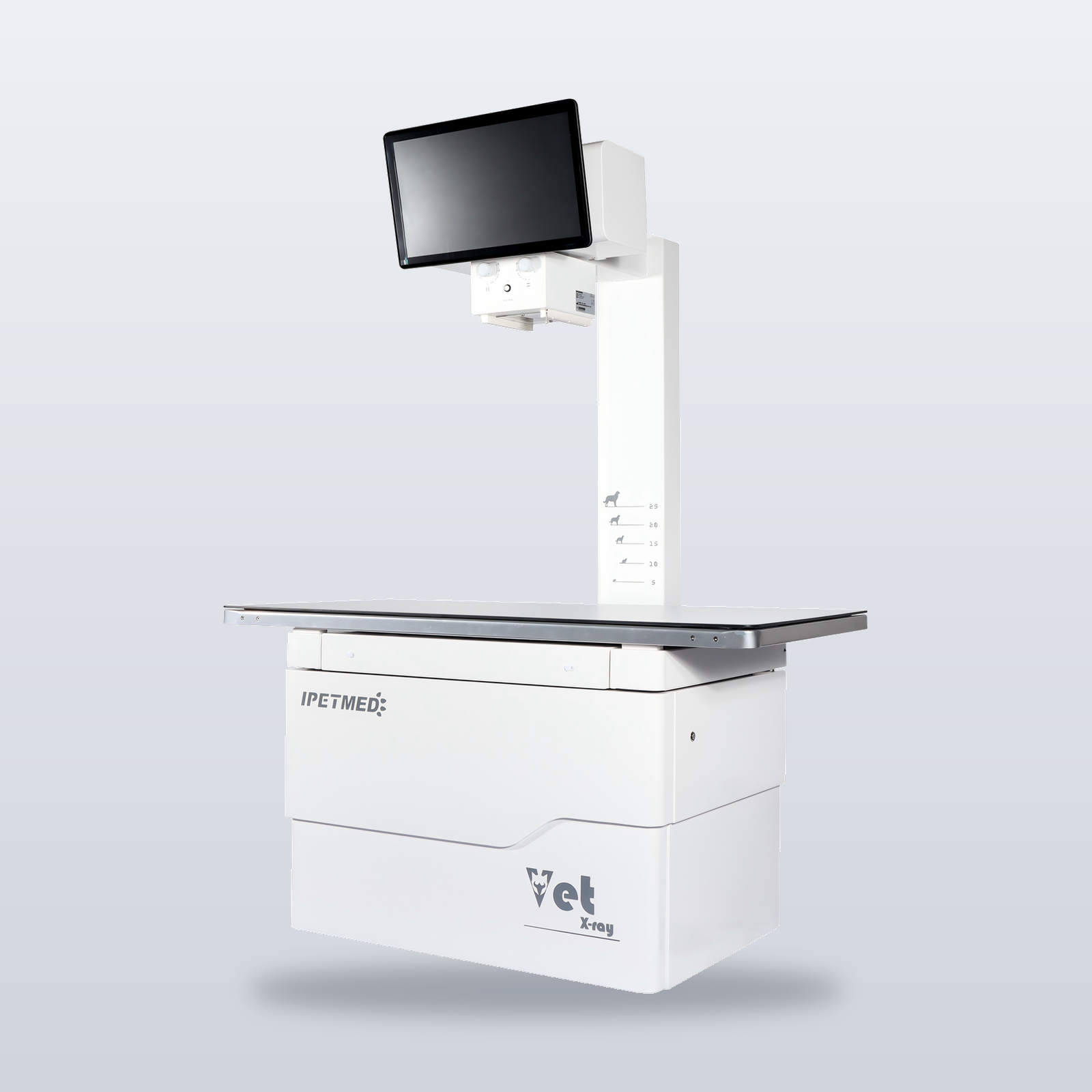
Dual floor-rail layout with auto-tracking chest stand and a separately tracked tube column. The tube head rotates ±180° for any beam orientation; the flat-panel detector mounts behind the JPI-1000 grid in the chest stand or on a fold-out table for recumbent imaging.
Power input
| Voltage | 380 V ±38 V |
| Frequency | 50 Hz ±1 Hz |
| Capacity | 135 kVA |
High-voltage generator (IWA-R80)
| Nominal power | 80 kW |
| Working frequency | 300 kHz |
| kV range | 40 – 150 kV |
| mA range | 10 – 1000 mA |
| Exposure time | 1 – 10000 ms |
| mAs range | 0.4 – 1000 mAs |
X-ray tube
| Target material | Rhenium-tungsten faced molybdenum |
| Target angle | 12° |
| Operating voltage | 40 – 150 kV |
| Anode input power | 65 kW (large focus) / 28 kW (small focus) |
| Anode heat capacity | 210 kJ (300 kHU) |
| Max heat dissipation | 1000 W (1410 HU/s) |
| Component heat capacity | 900 kJ (1250 kHU) |
| Max anode rotation | 3200 rpm |
| Focal spot | 1.0 mm (large) / 0.6 mm (small) |
| Inherent filtration | ≥ 1 mm Al |
Chest stand & tube column
| Chest stand vertical travel | 1500 mm (max 1785 mm, min 335 mm) |
| Grid focal length | 180 cm |
| Grid density | 103 LPI |
| Grid ratio | 10:1 |
| Tube rotation | ±180° |
| Tube column vertical travel | 1300 mm |
| Tube column horizontal travel | 2500 mm |
| Overall height | 2125 mm ±10 mm |
Flat-panel detector (IWA-FPD1717E)
| Scintillator | CsI |
| Detector technology | TFT + photodiode (a-Si) |
| Image size | 43 (H) × 43 (V) cm — 17×17″ |
| Pixel matrix | 3072 × 3072 |
| Pixel pitch | 139 µm |
| A/D conversion | 16 bit |
| Spatial resolution | ≥ 3.6 LP/mm |
| Energy range | 40 – 150 kVp |
Collimator & workstation
| Collimator illumination | ≥ 100 Lux average |
| Illumination timer | 30 s |
| Workstation | iVision Imaging System |
| DICOM | Patient registration, worklist retrieval, report writing, cloud platform |
| Image tools | Window level, zoom, rotation, mirror, ROI tools, sequence playback, image enhancement, label, calibration |
Software Configuration
| Acquisition workstation | In-house software including large-animal exposure parameter library |
| AI imaging assistant | Draft report generation; veterinarian reviews and signs off |
| DICOM | Patient registration, worklist retrieval, report writing, cloud platform |
Clinical Applications
- Equine thoracic, abdominal, and skeletal radiography in standing horses
- Equine distal-limb imaging with the tube column on horizontal beam
- Bovine and other large-ruminant radiography
- Large-breed canine and exotic-megafauna imaging in zoos and wildlife centres
- Teaching-hospital and research radiography for large animals
- Pre- and post-surgical imaging for equine orthopaedic procedures
Workflow & Installation
Three-phase 380 VAC ±38 V supply with 135 kVA capacity is required. The dual-rail floor plan, chest-stand vertical travel, and 2500 mm horizontal tube column travel demand site planning before installation; layout drawings are supplied during site survey. Operating environment per IWA installation manual; engineer-led installation, calibration, and training included.
Package Contents
- High-voltage generator (IWA-R80)
- X-ray tube assembly
- Manual collimator
- Chest stand (dual-rail, auto-tracking)
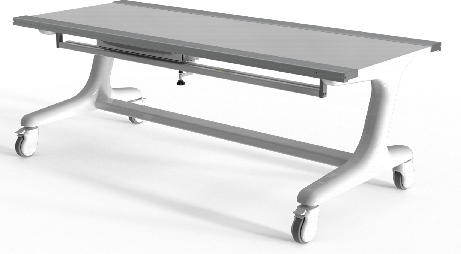
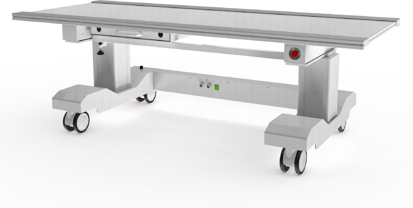
- Tube stand (X-ray table)
- JPI-1000 grid (10:1, 103 LPI, 180 cm focal length)
- 17×17″ CsI flat-panel detector (IWA-FPD1717E)
- iVision Imaging acquisition workstation
- Two complete lead protection sets (gown, hat, scarf — complimentary)
- iVision-1000X accessories
Downloads
Why Choose IWA-DR07
- Workflow efficiency — auto-tracking chest stand and ±180° tube rotation cover any standing-exam geometry without manual repositioning.
- Clinical accuracy — 1000 mA tube current at 80 kW shortens exposure to control motion blur in standing horses.
- Practice economics — dedicated large-animal layout removes the cost of patient relocation or off-site referral.
Warranty & Support
Two-year warranty including free parts and labour during the warranty period, excluding operator-caused damage and force majeure. Engineer-led installation, calibration, and operator training included. Two complete lead protection sets supplied at no charge.
FAQ
Q: What patients is iVision-1000X designed for?
Horses (full-body and distal-limb), cattle, large-breed dogs, and zoo or wildlife patients. The 80 kW generator and 1000 mA tube are sized for body masses where small-animal DR cannot deliver adequate exposure.
Q: Does it require three-phase power?
Yes. 380 VAC three-phase supply with 135 kVA capacity is required.
Q: Is the detector wireless?
The standard configuration uses a tethered 17×17″ CsI panel mounted in the chest stand bucky tray. Detector handling and mounting workflows are discussed during site planning.
Q: Can I image standing horses?
Yes. The dual-rail chest stand and ±180° rotating tube column are designed for standing equine thoracic, abdominal, and skeletal exams.
Q: Is the system DICOM PACS compatible?
Yes. iVision Imaging supports DICOM 3.0 Store, Print, Worklist, and cloud-platform reporting.
Reviews
There are no reviews yet.