
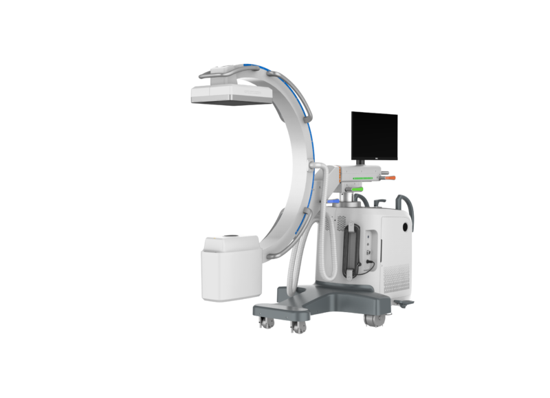
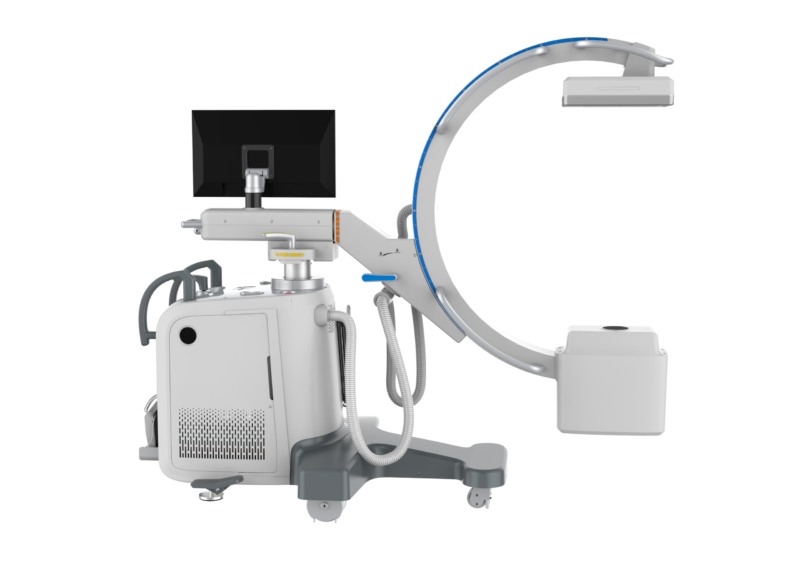
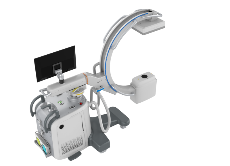
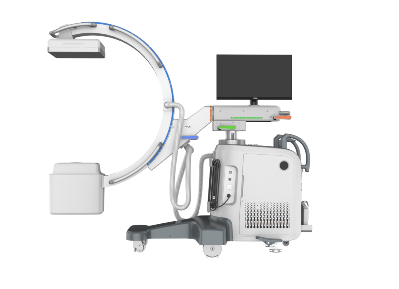
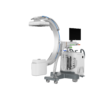
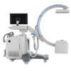
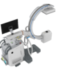
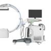
Real-time fluoroscopic guidance at the operating table — a mobile digital C-arm with a 5 kW generator and a flat-panel detector.
Product Overview
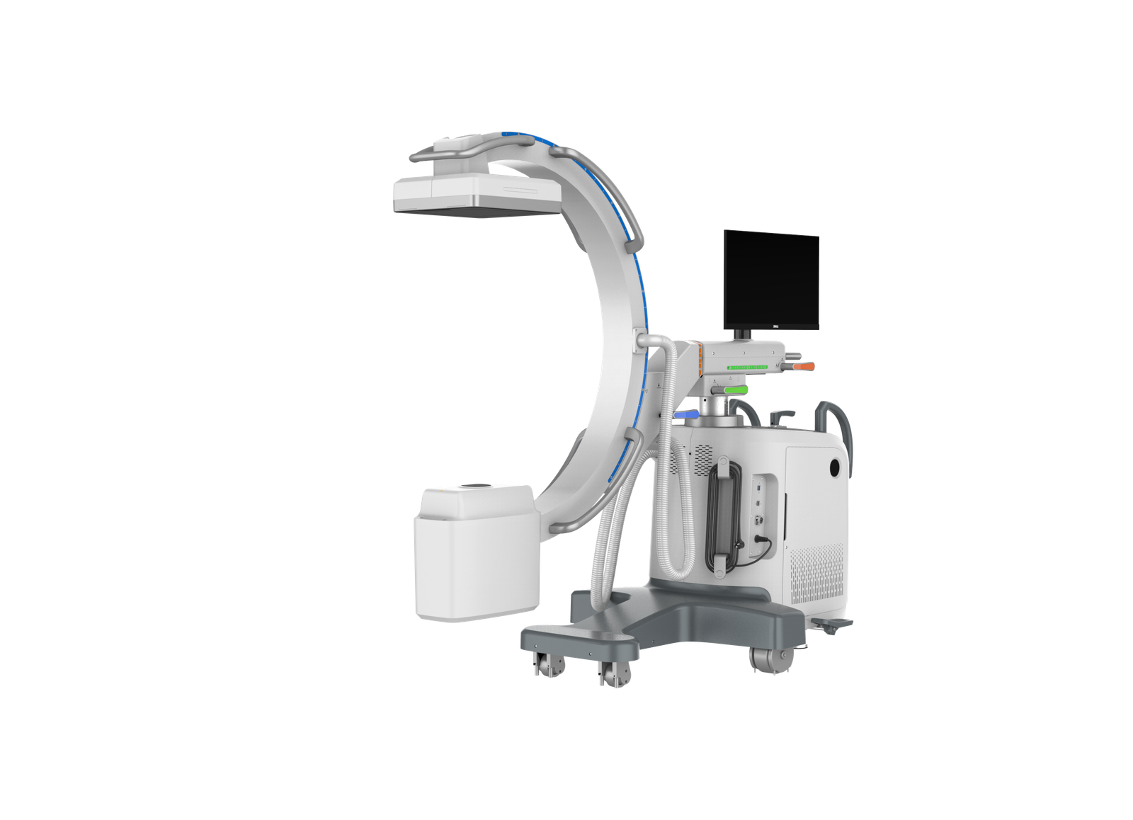
iPet-C5 is a compact, multi-functional digital C-arm fluoroscopy system that saves space in the practice and improves workflow. It provides radiological guidance and image display for diagnosis, interventional procedures and surgery in canine and feline patients.
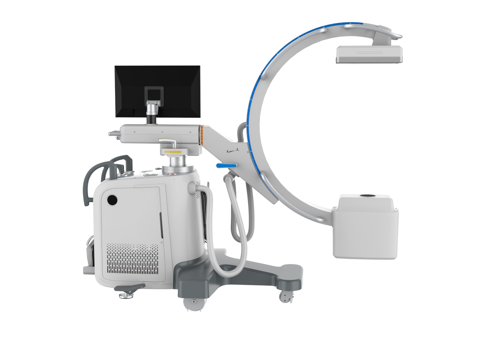
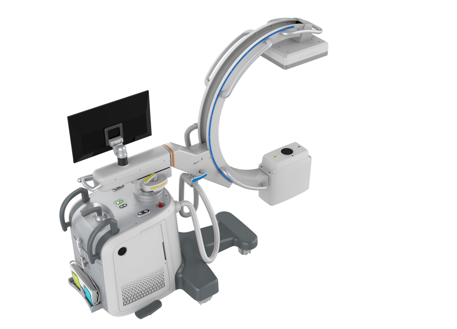
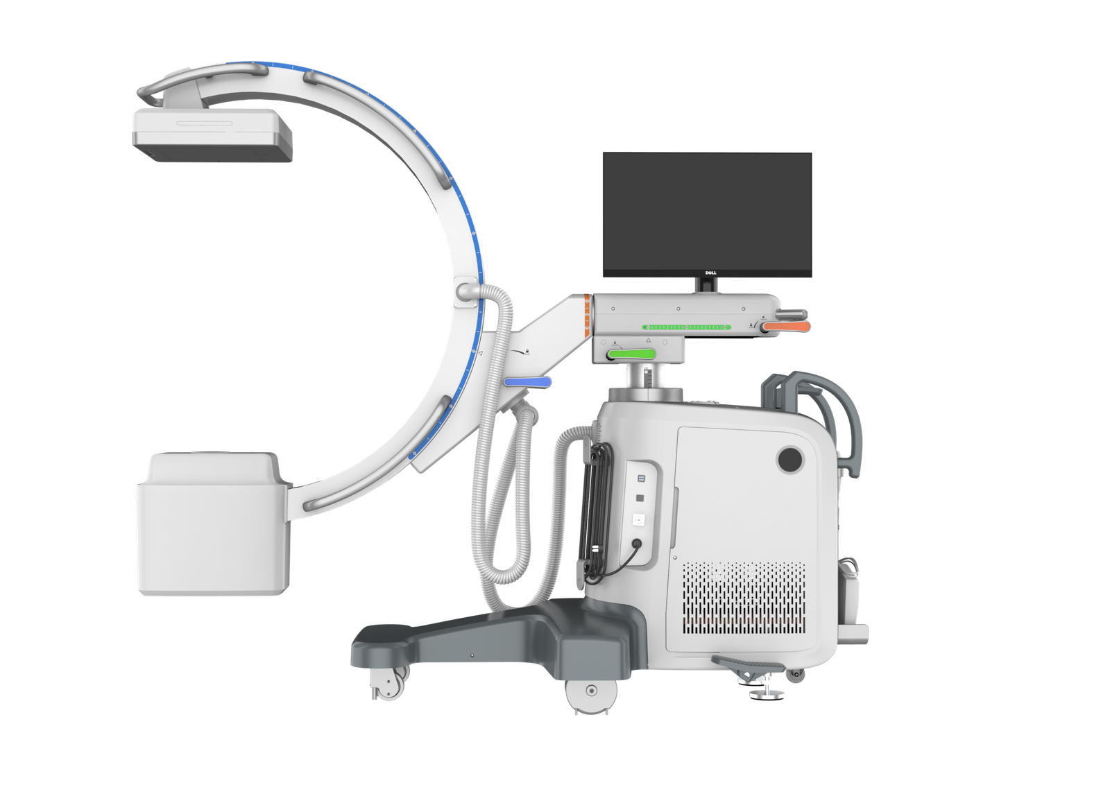
A 5 kW high-voltage generator with a rotating-anode tube (0.3 / 0.6 mm focal spots) is paired with an iRay Mercu0909F flat-panel detector. The C-arm slides, rotates, lifts, translates, swings and moves forward and backward, and the gantry monitor is operated entirely by touch, so the team repositions the C-arm rather than the patient.
Key Features
- Free-sliding C-arm — 150° orbital slide (+117° / -33°) reaches new projections without repositioning.
- 360° rotation — ±180° C-arm rotation for multi-angle views.
- Flexible positioning — 400 mm vertical movement, 200 mm lateral movement, ±15° swing and forward / backward movement to reach the desired position quickly.
- Three exposure modes — continuous 6.3 mA, pulsed 32 mA and digital spot 100 mA, with 10–1600 ms exposure time and 0.2–100 mAs.
- Flat-panel detector — iRay Mercu0909F, 210 × 210 mm effective area, 1024 × 1024 matrix, 205 µm pixel size, removable grid.
- Full-touch monitor — mode selection and parameter setting by touch at the gantry.
- Flexible mobility — the mobile unit transfers between rooms and positions at the table with little effort.
- Foot-switch acquisition — hands stay in the surgical field during image acquisition.
Product Gallery
Technical Specifications — Power, Generator and X-Ray Tube
| Model | iPet-C5 |
| Power input | 110 VAC / 220 VAC |
| Power | 5 kW |
| kV range | 40–125 kV |
| mA range | Continuous 6.3 mA · Pulsed 32 mA · Digital spot 100 mA |
| ms range | 10–1600 ms |
| mAs range | 0.2–100 mAs |
| Tube type | Rotating anode |
| Focal spot | 0.3 / 0.6 mm |
Technical Specifications — Detector
| Model | iRay Mercu0909F |
| Spatial resolution | 2.4 lp/mm |
| Pixel matrix | 1024 × 1024 |
| Pixel size | 205 µm |
| Removable grid | Yes |
| Effective imaging area | 210 × 210 mm |
| Data interface | 1G Ethernet |
Technical Specifications — Main Frame and C-Arm
| Main frame dimensions | 1886 × 1563 × 853 mm |
| Weight | Approx. 280 kg |
| SID | 1000 mm |
| Aperture | 784 mm |
| Arc depth | 640 mm |
| Orbital slide | 150° (+117° / -33°) |
| Rotation | 360° (±180°) |
| Swing | ±15° |
| Lateral movement | 200 mm |
| Vertical movement | 400 mm |
Clinical Applications
Workflow and Installation Notes
Operating workflow
- Prepare the operating table.
- Anaesthetise the patient.
- Apply radiation protection for all staff in the room.
- Move the C-arm close to the table.
- Position the imaging site in the C-arm field.
- Select the imaging mode and set exposure parameters.
- Check for collisions between the C-arm and the patient.
- Press the foot switch to acquire images.
Site preparation
The manufacturer’s reference layout places the C-arm in a 3000 × 4000 mm examination room next to a separate control room with a 1200 mm lead-glass window. Final layout and shielding follow local radiation-protection regulations.
| Power supply | Dedicated power box, 110 VAC / 220 VAC, with neutral and earth terminals; do not share with other equipment. Supply capacity and other electrical requirements follow the manufacturer’s installation requirements. |
| Room height | ≥ 2300 mm clear |
| Door width | ≥ 950 mm recommended |
| Floor | Level within 1.5 mm; concrete layer > 100 mm |
| Shielding | ≥ 3 mm Pb equivalent in the primary beam direction, 2 mm Pb equivalent elsewhere |
| Environment | 15–35 °C, 35–75 % RH; air-conditioning or dehumidification recommended; forced negative-pressure ventilation recommended |
| Cabling | 80 mm floor-level cable hole with lead maze; 150 × 100 mm cable trench |
| Workstation desk | Supplied by the practice |
Package Contents
- 1 × C-arm main unit with high-voltage generator and rotating-anode X-ray tube
- 1 × iRay Mercu0909F flat-panel detector with removable grid
- 1 × Gantry-mounted touch monitor
- 1 × Workstation
- 1 × Exposure foot switch
Downloads
Product brochure and site-preparation drawing are available on request — contact us.
Why Choose iPet-C5
- Workflow efficiency — the C-arm slides, rotates, lifts, translates and swings to the anatomy, and foot-switch acquisition keeps hands in the field; the patient stays on the table.
- Clinical accuracy — live fluoroscopy and digital spot images from a 2.4 lp/mm flat panel confirm reduction and implant position during the procedure.
- Space and mobility — a compact mobile unit that moves between the operating room and the examination room.
- Practice economics — one mobile unit covers surgical guidance, interventional work and diagnostic fluoroscopy.
Warranty and Support
Two-year warranty on the complete system (hardware and software), from the date of installation acceptance. Installation and operator training terms are confirmed in the written quotation — contact us for support in your region.
FAQ
Q: What exposure modes does iPet-C5 offer?
Continuous (6.3 mA), pulsed (32 mA) and digital spot (100 mA), within a 40–125 kV range, 10–1600 ms exposure time and 0.2–100 mAs.
Q: Which detector is fitted?
An iRay Mercu0909F flat panel: 210 × 210 mm effective imaging area, 1024 × 1024 matrix, 205 µm pixels, 2.4 lp/mm, removable grid and 1G Ethernet interface.
Q: How far can the C-arm move?
150° orbital slide, ±180° rotation, ±15° swing, 400 mm vertical and 200 mm lateral movement, plus forward / backward movement, with 1000 mm SID, 784 mm aperture and 640 mm arc depth.
Q: What room does it need?
A shielded room (3 mm Pb primary direction, 2 mm Pb elsewhere) at least 2300 mm high, a door of at least 950 mm, and a dedicated 110 VAC / 220 VAC power box. The reference layout uses a 3000 × 4000 mm examination room.
Q: What is the warranty?
Two years on the complete system, from the date of installation acceptance.
Reviews
There are no reviews yet.