
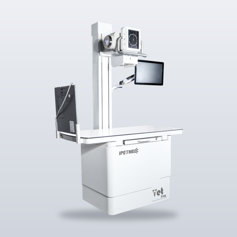
The full spine of a large-breed canine in one exposure — iPet-1724, the built-to-order large-format system of the iPet AI-DR line.
Product Overview
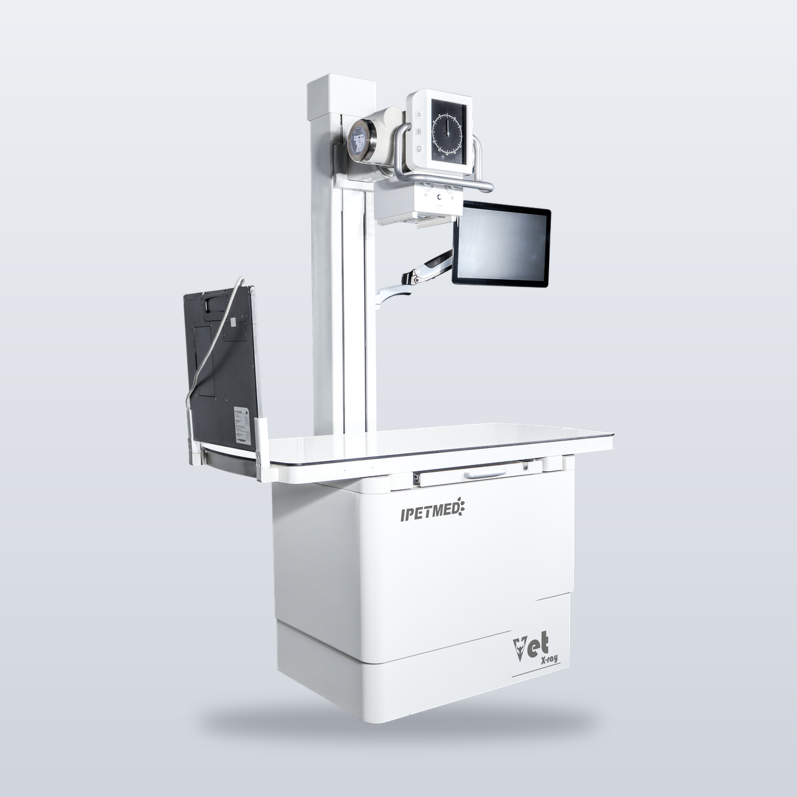
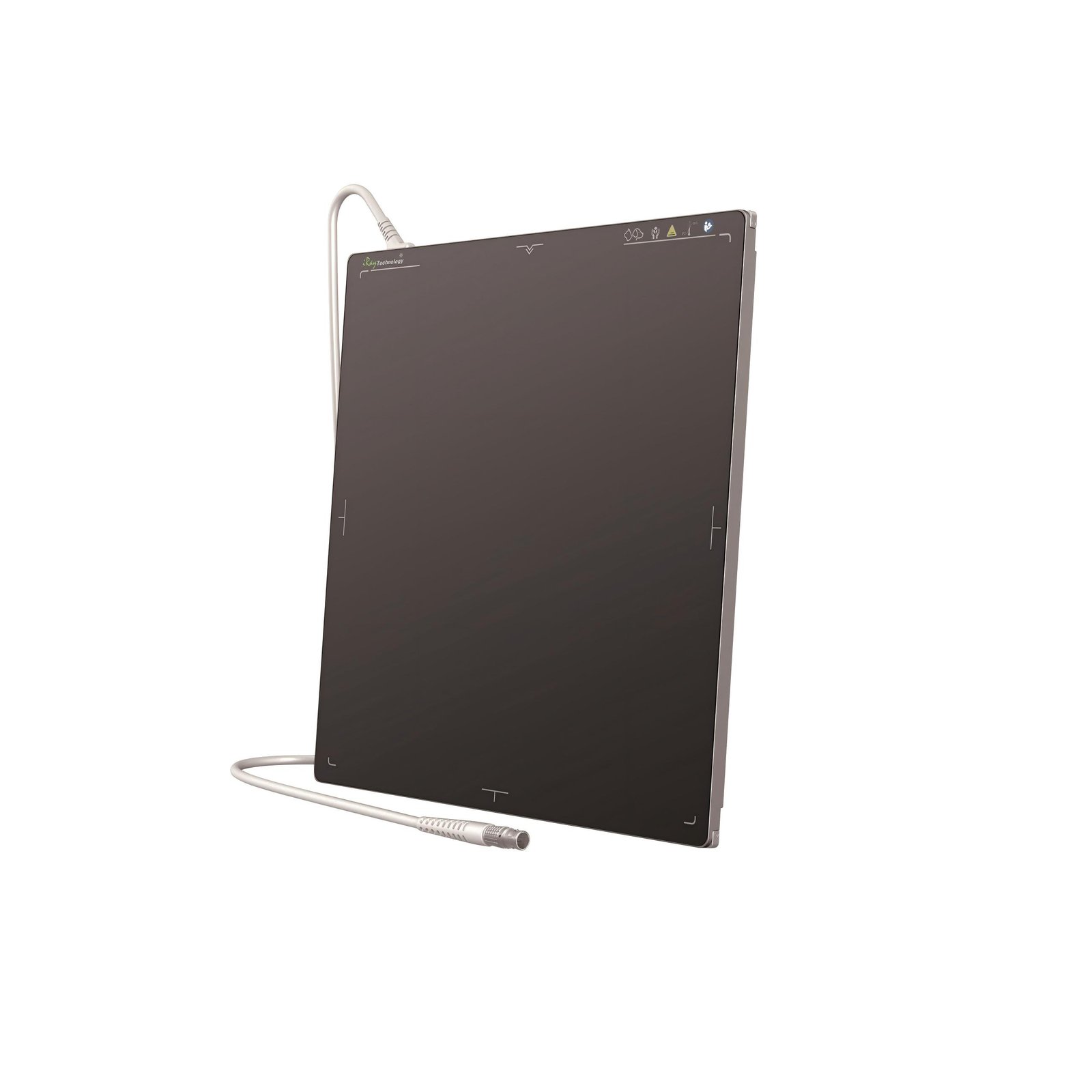
A full-spine study of a Great Dane on a 43 × 43 cm panel takes three segments and three stitched images; when the stitch line falls across the lesion, the study has to be repeated. iPet-1724 enlarges the imaging area to 17 × 24 inches (43 × 61 cm), so the full canine spine and both hip joints are captured in one exposure — one exposure fewer, one restraint fewer, no stitching error. It is built to order on the iPet Pro platform: elevating and rotating gantry, 40 kW high-frequency generator, and an original iRay 1724 flat panel, with configuration and lead time confirmed per order.
Key Features
- 17 × 24″ large format — full spine and both hips in a single exposure.
- Fewer exposures, less restraint, no stitching error.
- 40 kW high-frequency generator — penetration headroom for large, thick body regions.
- Original iRay 1724 flat panel — serial number verifiable.
- Pro platform heritage — elevating and rotating gantry for standing and natural-position views.
- Same in-house software stack as the flagship — acquisition workstation, AI imaging assistant, cloud imaging.
- DICOM worklist — order straight to DR — exam orders from practice software arrive at the workstation automatically, with no second entry of patient data.
System Specifications (Confirmed per Order)
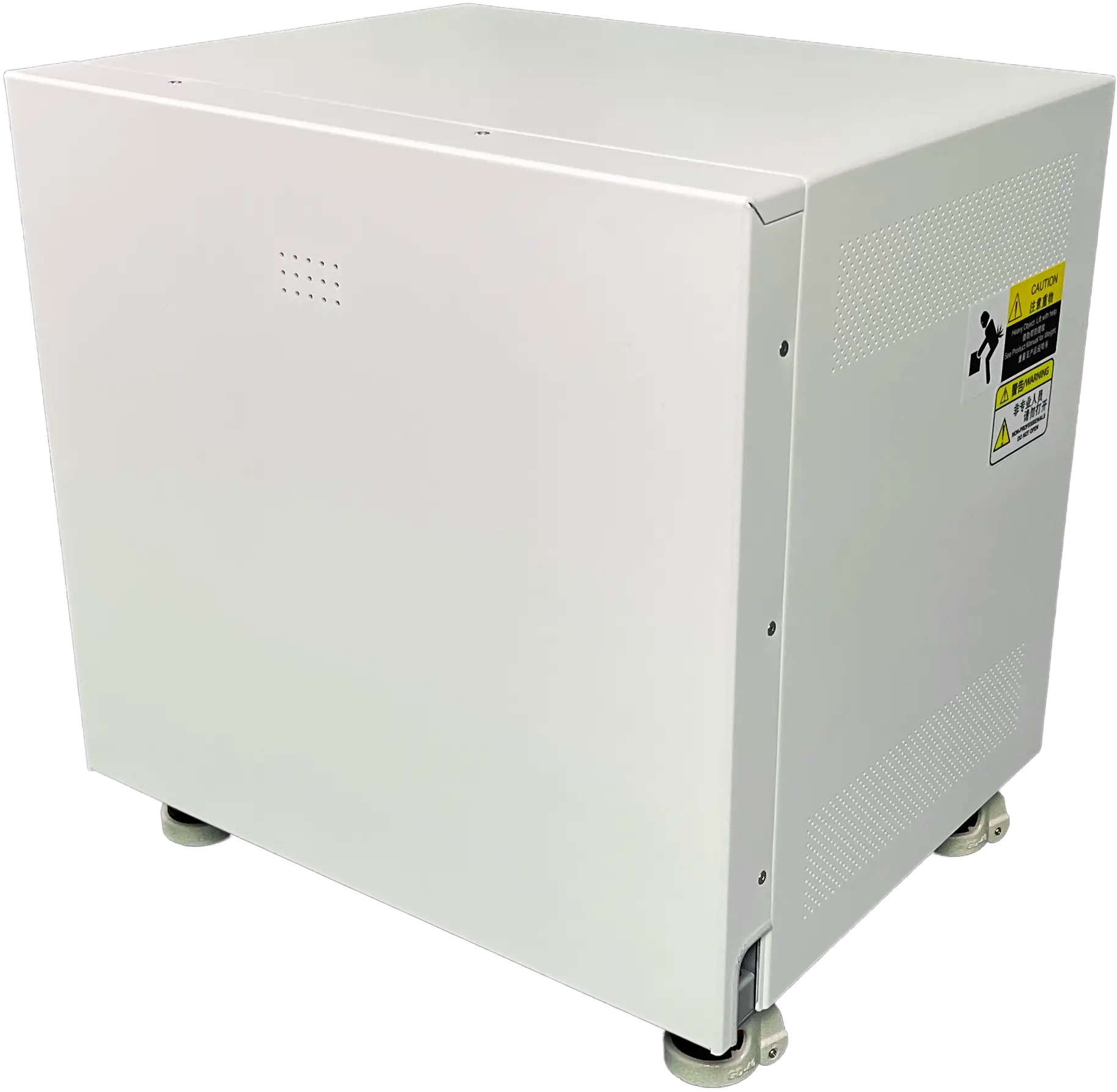
| High-voltage generator | 40 kW high-frequency (second-generation veterinary) |
| X-ray tube | Canon (model confirmed per order) |
| Gantry | Elevating and rotating gantry (Pro platform): height travel, 90° rotation for horizontal-beam projection |
| Flat-panel detector | Original iRay 1724, 17 × 24 inches (approx. 43 × 61 cm), serial number verifiable; 139 µm, directly deposited CsI, AED (exact parameters per final technical documentation) |
| Workstation | Industrial PC with touchscreen |
| Power supply | 220 V (power requirements confirmed with the proposal) |
Software Configuration
| Acquisition workstation | In-house software, body-size automatic exposure |
| AI imaging assistant | Draft report generation; veterinarian reviews and signs off |
| DICOM / cloud | DICOM 3.0 Worklist; cloud image storage supported |
Clinical Applications
- Full-spine and bilateral hip studies of large-breed canine patients in one exposure
- Large-field thoracic and abdominal radiography of giant-breed canine patients
- Routine radiography — the larger panel covers more anatomy per view
Build-to-Order Notes
Built to order: panel orientation (landscape or portrait priority), table dimensions and related details are confirmed with each order. Lead time follows the production schedule confirmed at order.
Downloads / Documentation
Specification sheet available on request.
Get a Proposal
Built to order, no list price. Contact our sales team to confirm configuration and lead time. For standard 43 × 43 cm imaging, see the iPet-400Pro static flagship.
Warranty & Support
2-year warranty on the complete system. The flat panel carries the original manufacturer warranty and ships with a serial-number verification card. On-site installation, calibration, and operator training by our engineers.
FAQ
Q: How much larger is it than a 43 × 43 cm panel?
The active imaging area grows from 43 × 43 cm to approx. 43 × 61 cm — full-spine and bilateral hip studies of large-breed canine patients move from segmented acquisition and stitching to a single exposure.
Q: Is the panel original?
Yes. An original iRay 1724 flat panel; the serial number is verifiable through iRay’s official channel, and a verification card ships with every system.
Q: Why is there no list price?
This is a built-to-order system: panel orientation, table dimensions, and tube configuration differ per order, so pricing is issued with the quotation.
Q: What is the lead time?
Built to order; lead time follows the production schedule confirmed at order. Contact our sales team for an assessment.

Reviews
There are no reviews yet.