
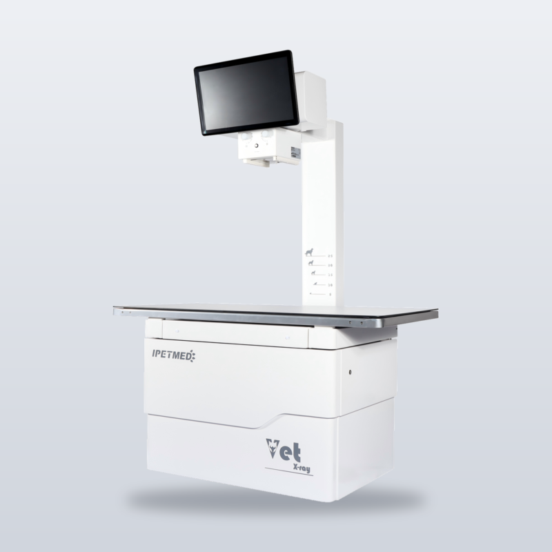
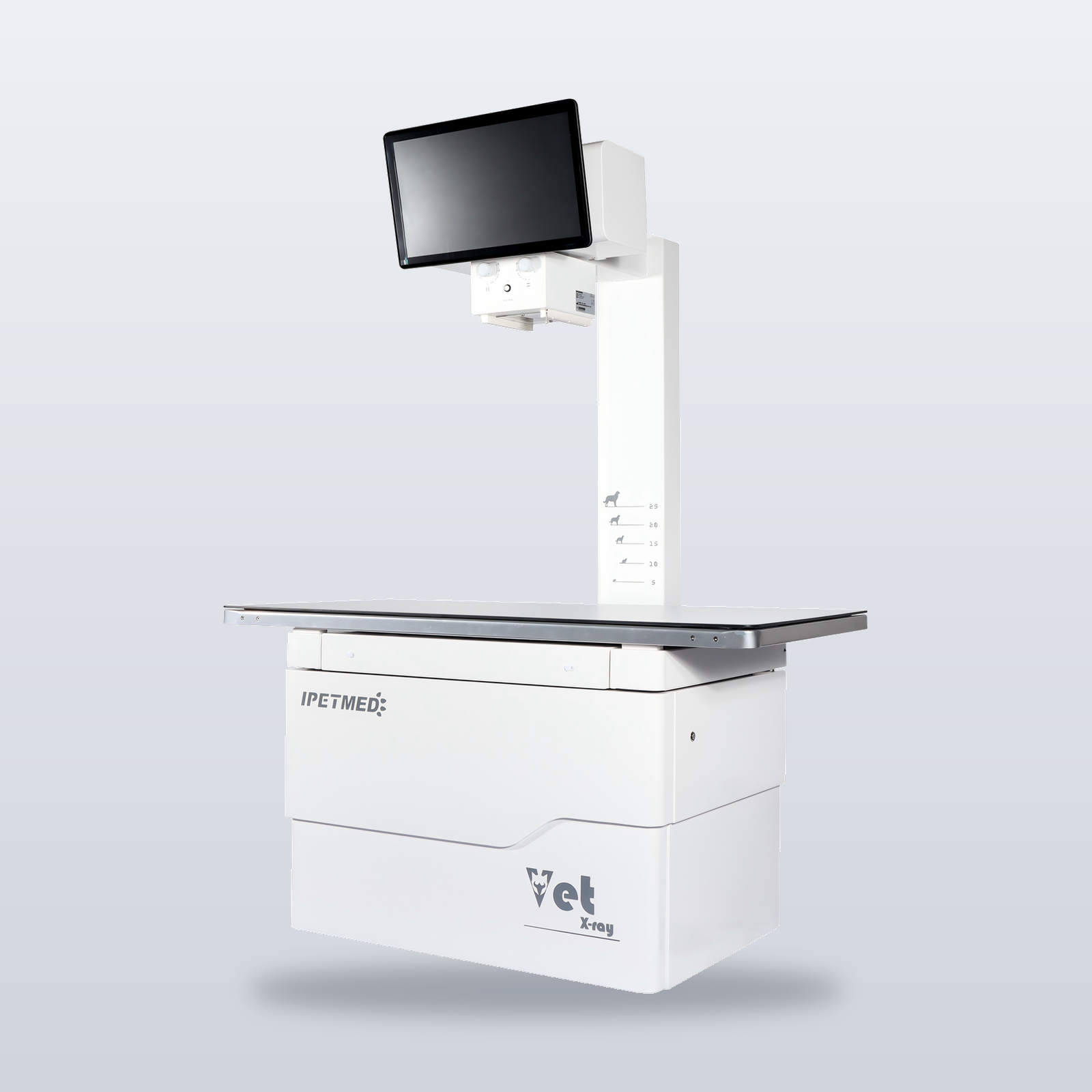
The iPet-400AI X-ray platform without the detector and workstation — a direct replacement for an ageing or failed veterinary X-ray unit.
Product Overview
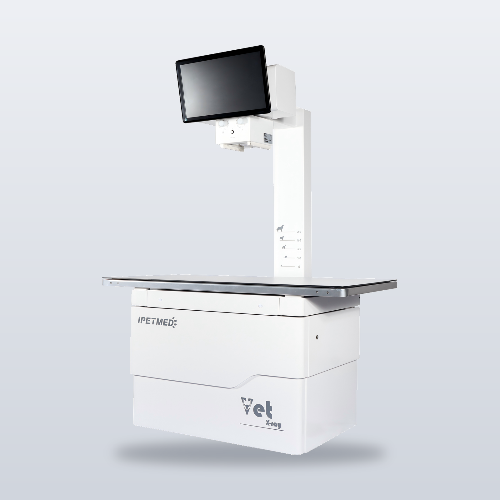
iPet-X is the X-ray unit of the iPet-400AI platform. It keeps the 32 kW high-frequency generator, Canon E7239X rotating-anode tube, LED collimator, 1400 × 700 mm four-way floating table and fixed tube column, and removes the flat-panel detector, acquisition workstation and imaging software.
It is built for practices whose X-ray machine is ageing or out of service and needs replacing. Image receptor equipment is not included; it is provided by the user and is outside the scope of supply.
Key Features
- 32 kW high-frequency generator — 40 – 125 kV, 10 – 400 mA, 1 – 10000 ms; kV-mA-ms and kV-mAs exposure techniques.
- Canon E7239X rotating-anode tube — 1.0 / 2.0 mm dual focal spot, 140 kHU anode heat capacity.
- Four-way floating table — 1400 × 700 mm phenolic resin top, 200 kg load rating, foot-pedal release for single-handed positioning.
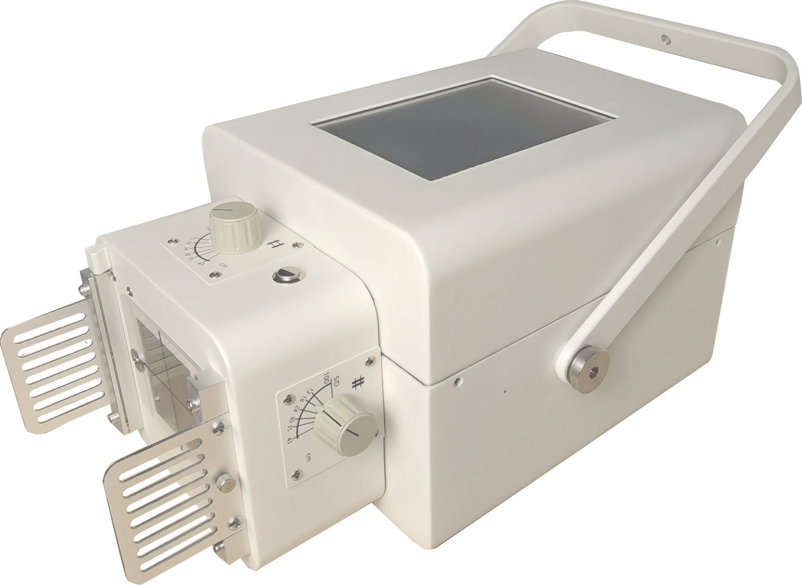
- LED collimator — 43 × 43 cm light field at 1 m SID, 1.0 mm Al inherent filtration at 75 kV.
- Separate exposure control panel — kV, mA, ms / mAs and focal spot are set on a dedicated control panel.
- Same platform as iPet-400AI — the same generator, tube, collimator and table as the AI-DR system.
Product Gallery
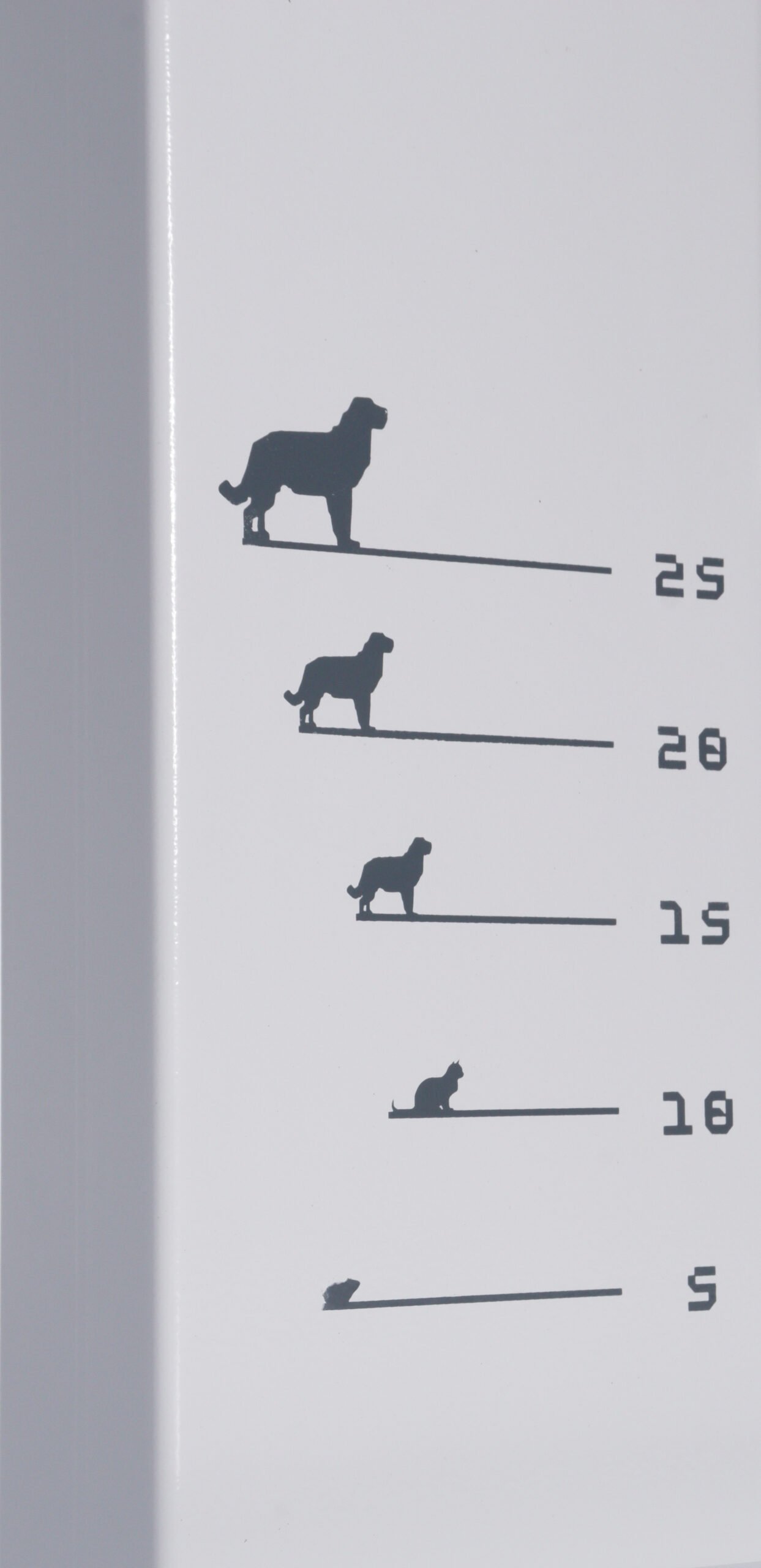
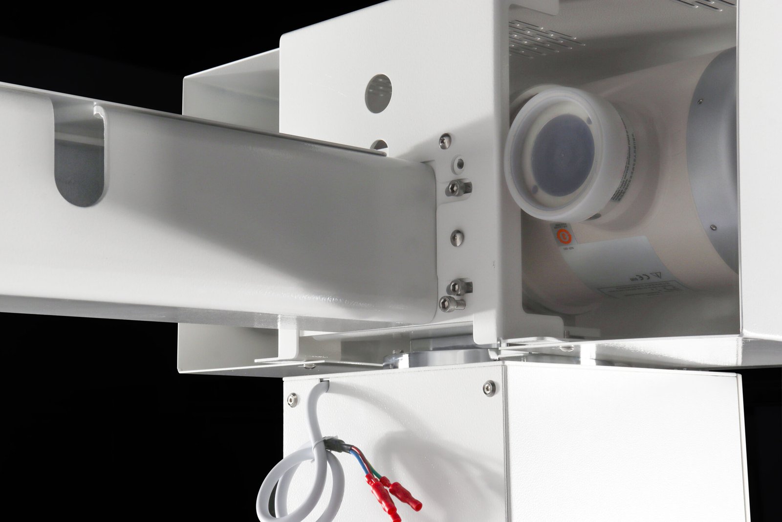
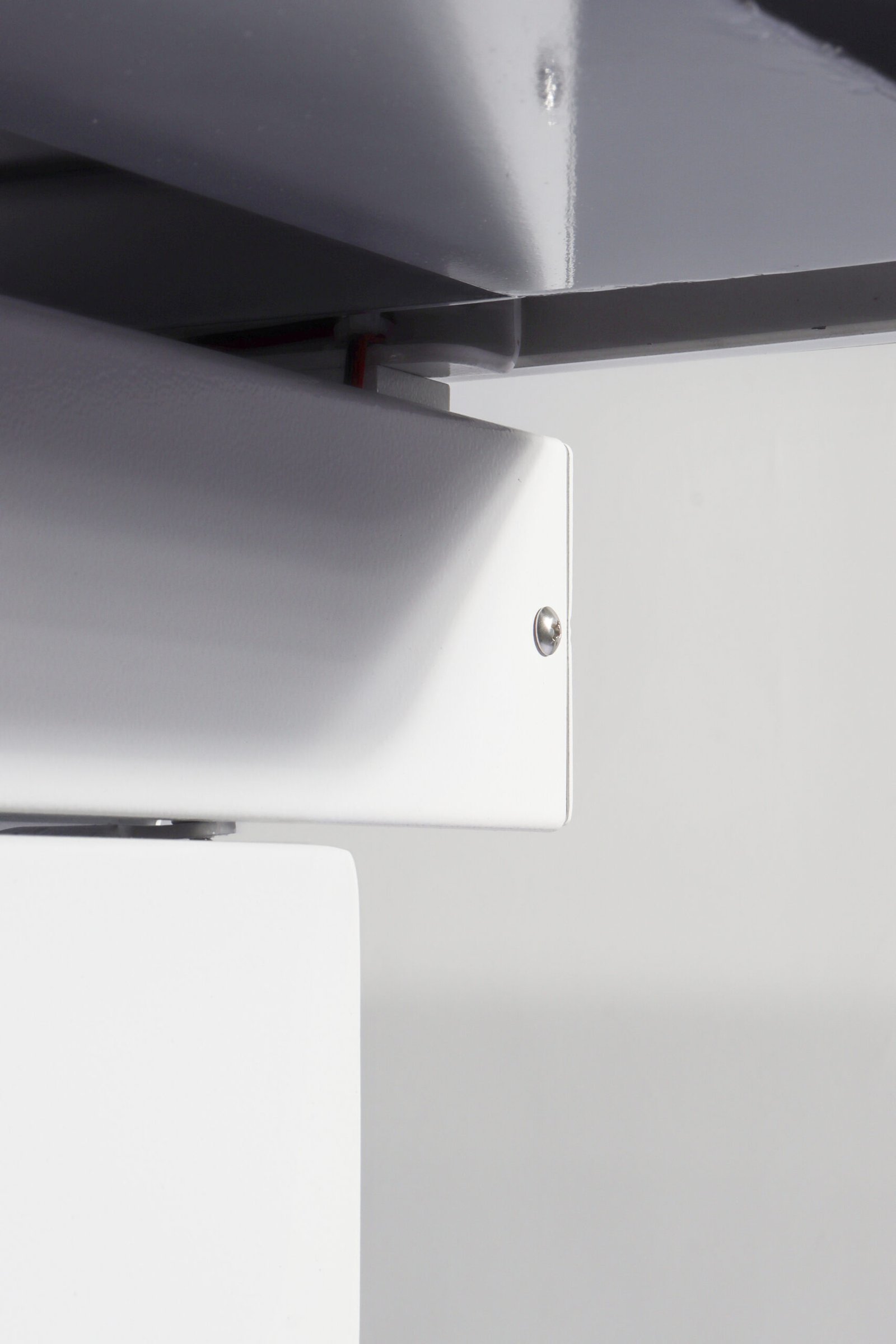
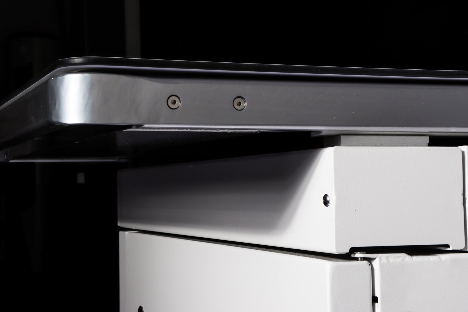
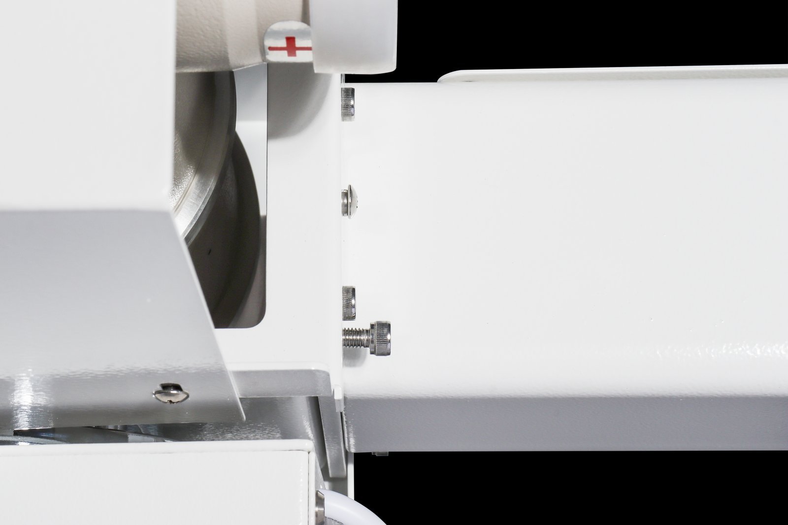
Detail views of the components iPet-X shares with the iPet-400AI platform.
System Specifications
| High-voltage generator | 32 kW high-frequency (veterinary), single phase 110/220 VAC |
| kV range | 40 – 125 kV |
| mA range | 10 – 400 mA |
| Exposure time | 1 – 10000 ms |
| Exposure technique | kV-mA-ms / kV-mAs |
| X-ray tube | Canon E7239X rotating anode |
| Tube max voltage | 125 kV |
| Focal spot | 1.0 / 2.0 mm (dual) |
| Anode input power | 21 kW (small focus) / 42.5 kW (large focus), 0.1 s |
| Anode heat capacity | 140 kHU |
| Patient table | 1400 × 700 mm, phenolic resin top, four-way float (X 75 ±10 mm / Y 200 ±10 mm) |
| Table height / load rating | 780 mm / 200 kg |
| Column height / SID | ≤ 2000 mm / ≥ 1000 mm |
| Collimator | LED light source, 1.0 mm Al filtration at 75 kV, 43 × 43 cm light field at 1 m SID |
| Exposure control | Separate control panel; two-stage hand switch and foot switch |
| Power supply | Single-phase 220 V ±10 %, 50 Hz, protective earth required; current rating [TBC] |
| Overall dimensions / weight | [TBC] |
| Operating environment | 10 – 38 °C, 20 – 75 % RH, 62 – 106 kPa |
Image Receptor
iPet-X does not include image receptor equipment. The receptor is provided by the user and is outside the scope of supply.
Clinical Applications
Replacement of an ageing or failed X-ray unit, used with the practice’s own image receptor. General canine and feline radiography — orthopaedic, thoracic and abdominal studies, pre-anaesthetic evaluation and post-operative rechecks — and routine imaging of small and medium exotic species.
For an integrated digital system, see iPet-400AI. For dynamic studies (barium swallow, airway evaluation), see the iSmart-32Pro dynamic DR system.
Workflow & Installation
- Exposure control — kV, mA, ms / mAs and focal spot are set on a separate control panel; exposure is triggered with the two-stage hand switch or the foot switch.
- Exposure charts — iPet-X has no body-size automatic exposure or protocol library. Build an exposure chart from the previous unit’s settings and test exposures, recorded against SID and receptor type.
- Image receptor — not included; provided by the user and outside the scope of supply.
- Power — single-phase 220 V mains with protective earth; no three-phase supply required.
- Installation — the room must accommodate the 1.4 m table and fixed tube column. Engineer-led installation and operator training are included.
Package Contents
- 32 kW high-frequency generator + control box
- Separate exposure control panel + two-stage hand switch
- X-ray tube assembly (Canon E7239X) + high-voltage cables
- LED collimator
- Patient table (1400 × 700 mm) with four-way floating top and fixed tube column
- Foot switch
- V-shape positioning set (1 large + 1 small)
- Lead protection set: gown, hat, scarf, glasses, gloves
- Power cable, operation manual, certificate of conformity
Flat-panel detector, acquisition workstation and imaging software are not included. Image receptor equipment is provided by the user and is outside the scope of supply.
Downloads
Documentation for iPet-X is available on request.
Why Choose iPet-X
- Workflow efficiency — replaces the X-ray unit alone, with exposure parameters set on a separate control panel.
- Clinical accuracy — 32 kW high-frequency output and a Canon rotating-anode tube give consistent exposures across body sizes.
- Practice economics — no detector or workstation included, so the practice pays only for the X-ray unit.
Warranty & Support
Two-year warranty covering the generator, tube, collimator and table. Extended-warranty service is available. Engineer-led on-site installation and operator training included.
FAQ
Q: Does iPet-X produce digital images on its own?
No. iPet-X is the X-ray unit only. Image receptor equipment is not included; it is provided by the user and is outside the scope of supply.
Q: Where are exposure parameters set?
On a separate control panel: kV, mA, ms or mAs, and focal spot. Exposure is triggered by the hand switch or foot switch.
Q: How does iPet-X differ from iPet-400AI?
Generator, tube, collimator and table are the same. iPet-X has no flat-panel detector, acquisition workstation, imaging software, AI imaging assistant or DICOM functions.
Q: Does the system require three-phase power?
No. Single-phase 220 V mains with protective earth is sufficient.
Reviews
There are no reviews yet.