
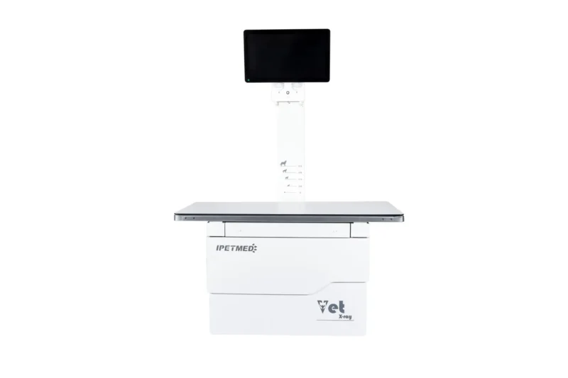
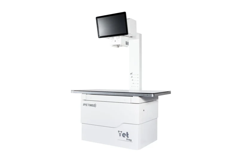
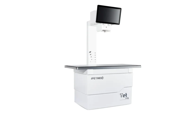
Veterinary digital radiography system built around a 32 kW high-frequency generator and 17×17″ flat-panel detector for daily small-animal practice.
Product Overview
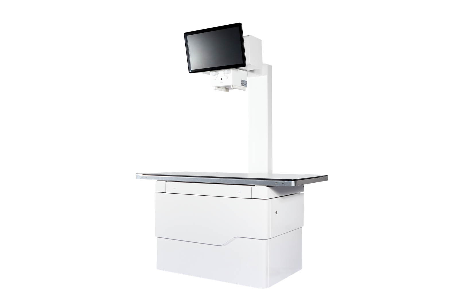
IWA-DR01 iPet-400 Veterinary DR X-ray Imaging System is the classical-configuration entry to the IWA small-animal DR line. The system pairs a 32 kW high-frequency generator and a Canon E7239X rotating-anode X-ray tube with a 43×43 cm caesium-iodide flat-panel detector and the iVetXray workstation, delivering one-second image acquisition for routine canine, feline, and exotic patients.
Designed as a fixed-room installation, IWA-DR01 covers the full range of general radiography — thorax, abdomen, skeletal, dental — in a configuration optimised for clinics that need reliable, high-throughput static imaging without the cost of dynamic fluoroscopy.
Key Features
- 32 kW high-frequency generator covering 40–150 kV / 10–400 mA / 1–10000 ms for full small-animal anatomy.
- Canon E7239X rotating-anode X-ray tube with dual focus 1.0 / 2.0 mm and 140 kHU anode heat capacity for sustained throughput.
- 17×17″ CsI flat-panel detector, 3072×3072 matrix, 139 µm pitch, 16-bit A/D, 3.6 LP/mm spatial resolution.
- One-second image acquisition via GigE interface — preview on the workstation in under a second after exposure.
- iVetXray acquisition software with built-in veterinary measurement tools (VHS, TPLO, HIP, TTA).
- Dual-display workstation: 23.8″ diagnostic monitor plus 21.5″ tableside touchscreen.
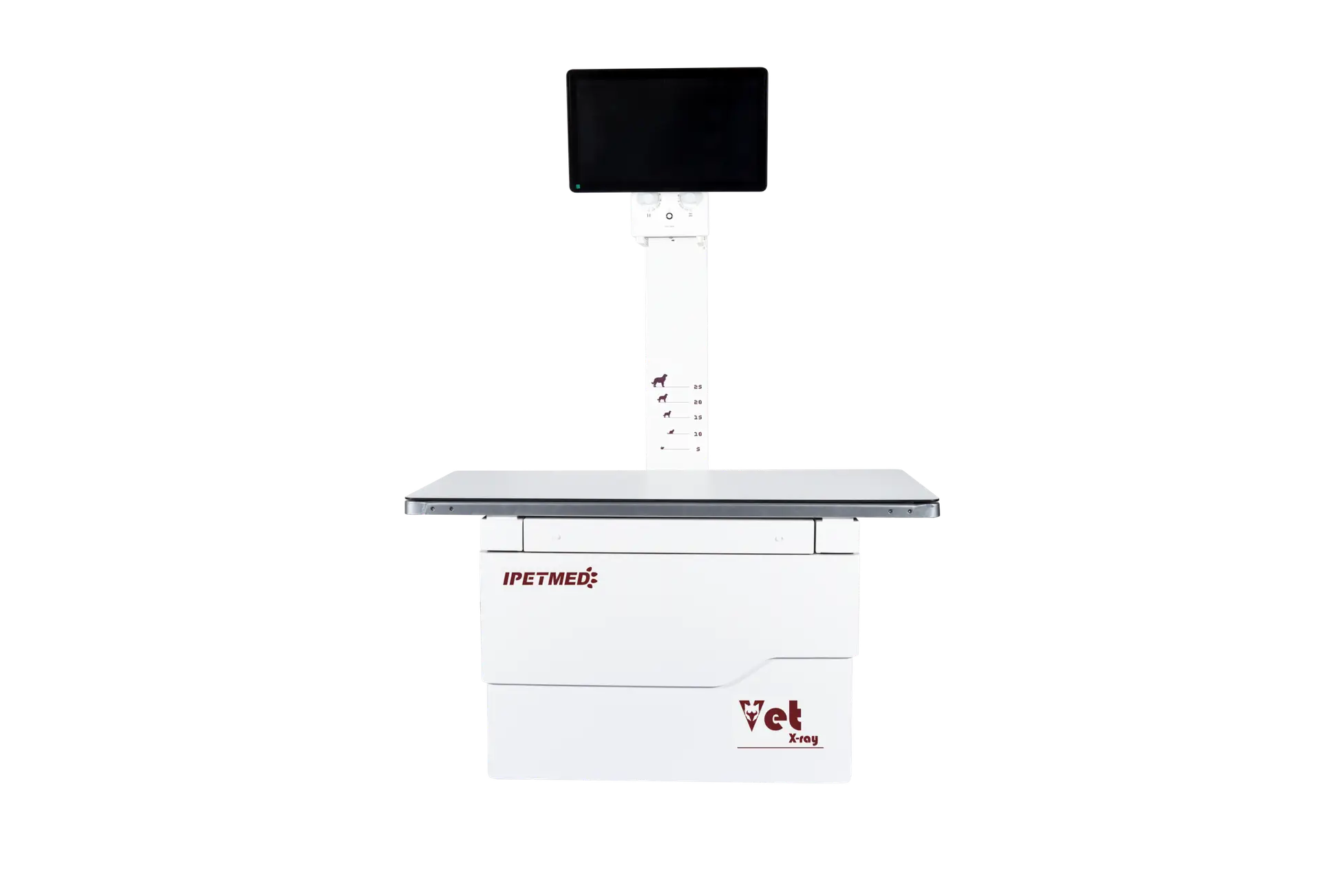
- Veterinary patient table 1400×700 mm, phenolic-resin top, ±75 mm longitudinal and ±200 mm lateral float.
- LED collimator with 1.0 mm Al inherent filtration at 75 kV; manual field setting.
- DICOM 3.0 compliant workstation with MiniPACS image viewer, print, and PACS integration.
Product Gallery
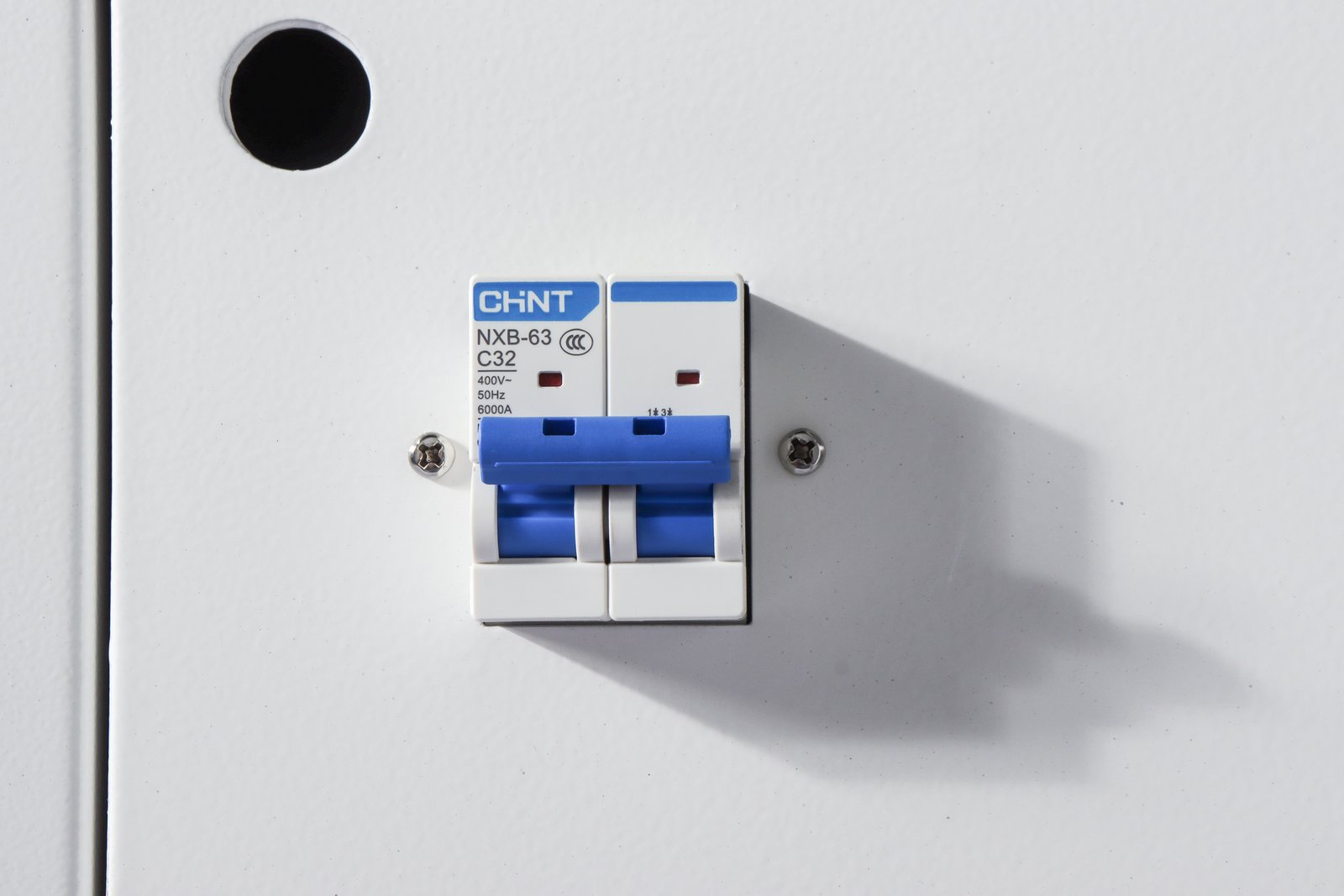
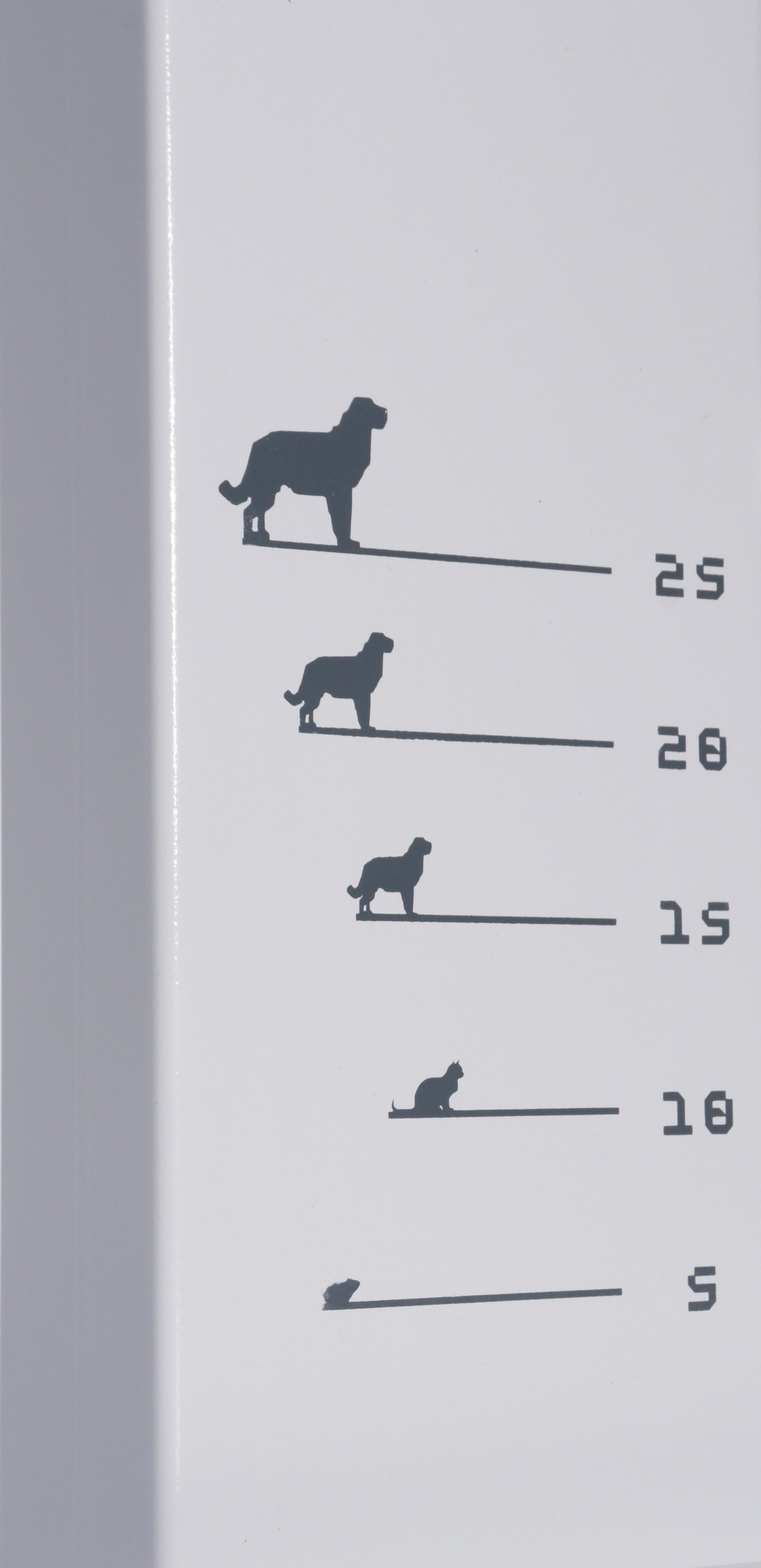
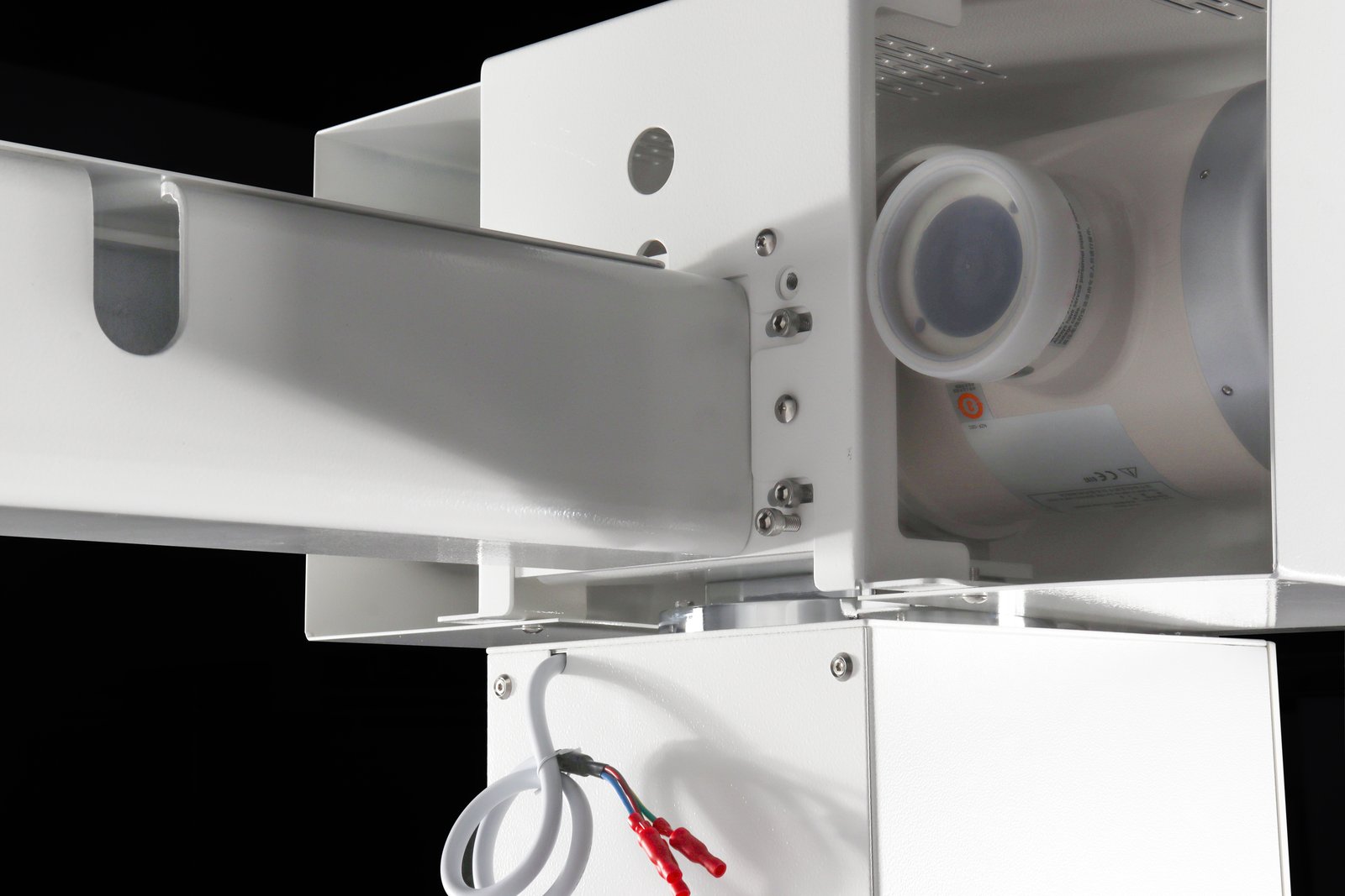
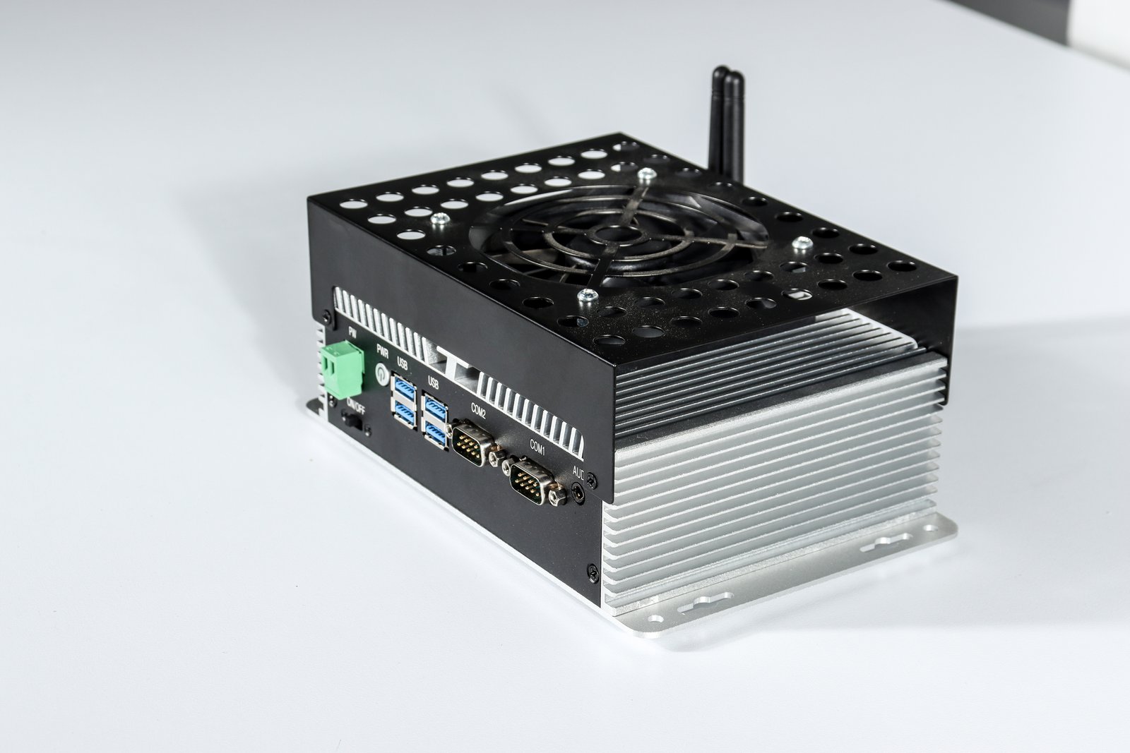
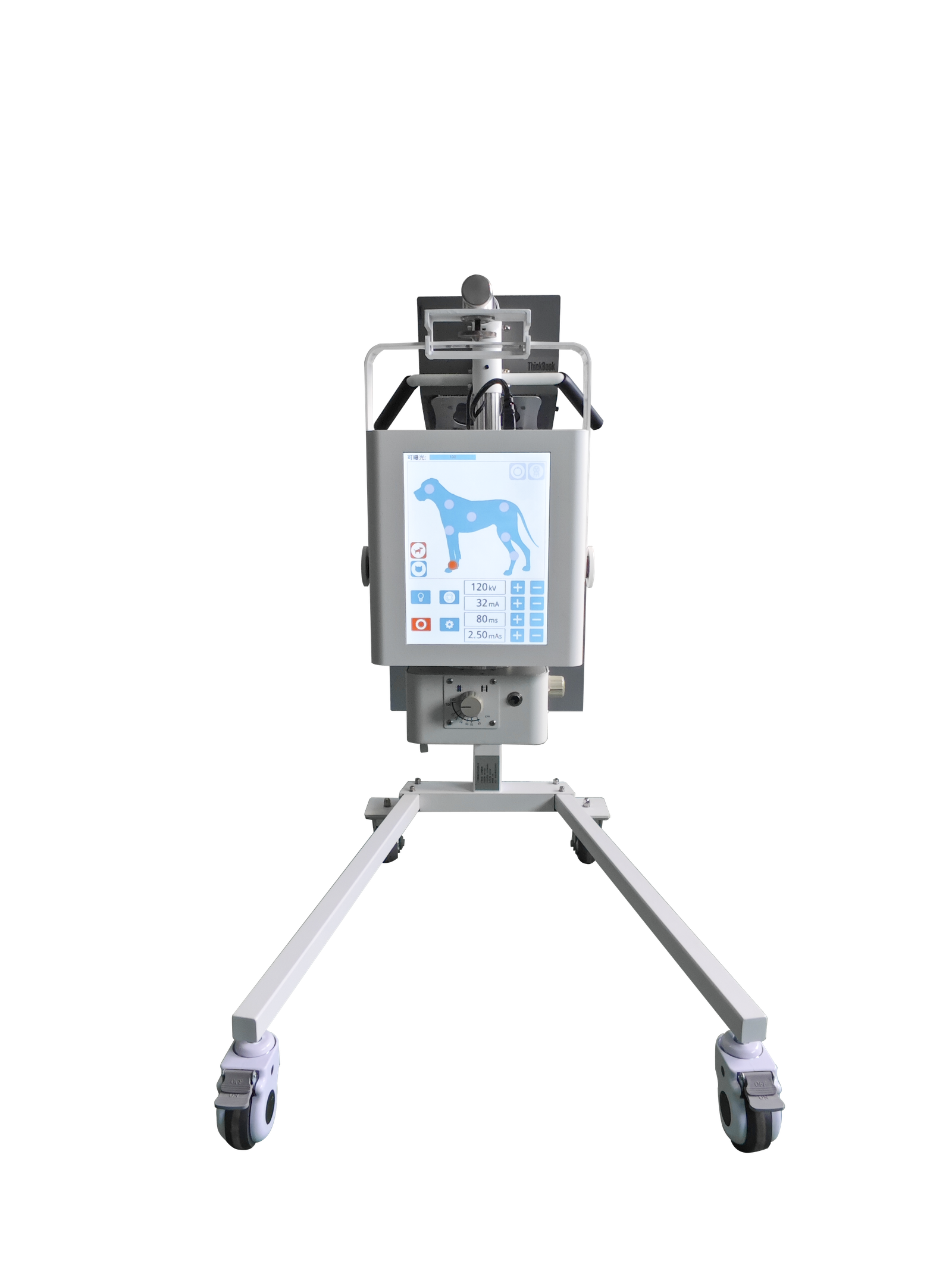
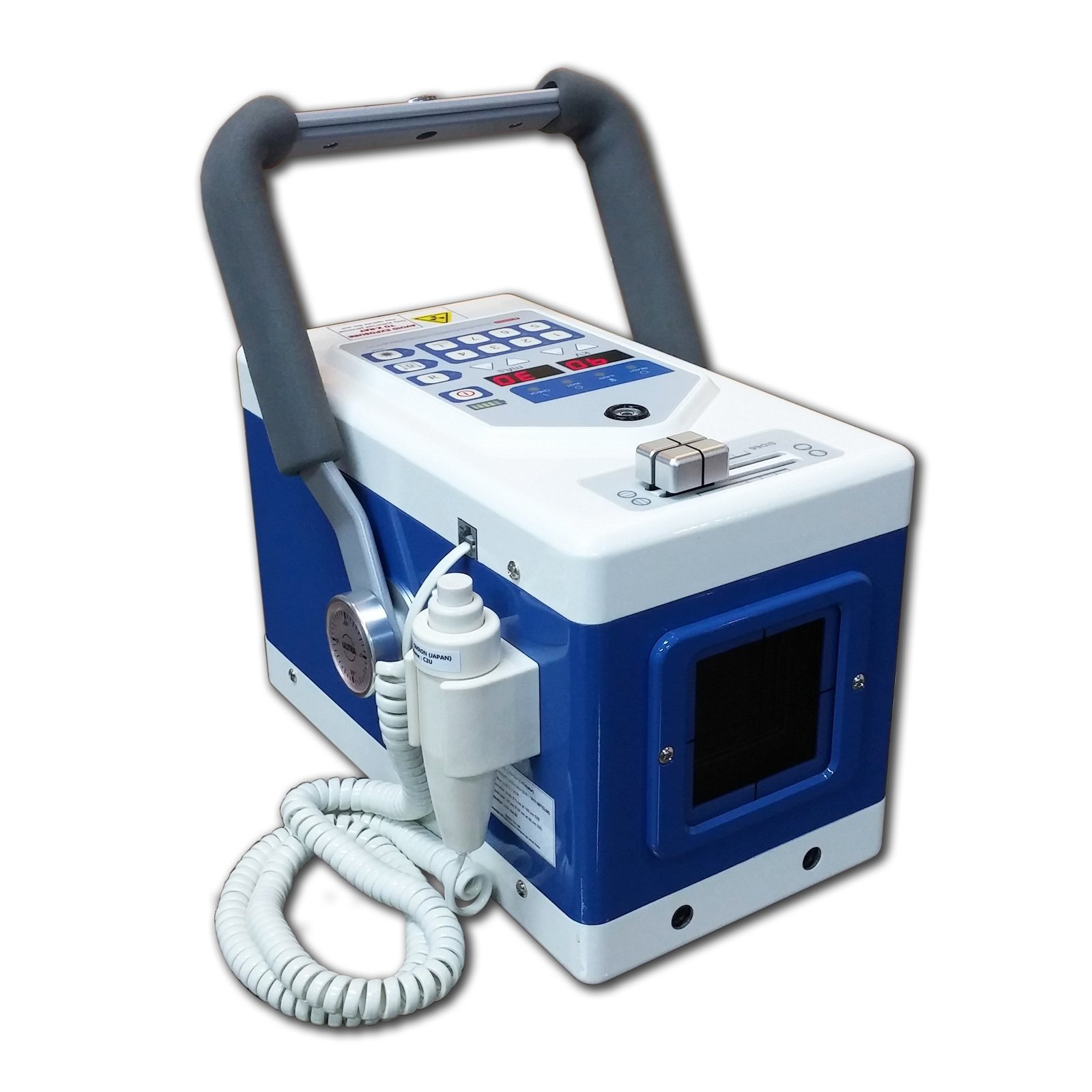
Detail views of the gantry, X-ray tube head, four-way floating patient table, LED collimator, and operator workstation.
Technical Specifications
High-voltage generator
| Output power | 32 kW |
| kV range | 40 – 150 kV |
| mA range | 10 – 400 mA |
| mAs range | 0.1 – 100 mAs |
| Exposure time | 1 – 10000 ms |
| Power input | Single phase, 110/220 VAC ±10% |
X-ray tube (Canon E7239X)
| Max voltage | 125 kV |
| Focal spot | 1.0 / 2.0 mm (dual) |
| Anode input power | 21 kW (small) / 42.5 kW (large) |
| Anode heat capacity | 140 kHU |
Flat-panel detector
| Technology | a-Si TFT, CsI scintillator |
| Image size | 43 × 43 cm (17×17″) |
| Pixel matrix | 3072 × 3072 |
| Pixel pitch | 139 µm |
| A/D conversion | 16 bit |
| Spatial resolution | 3.6 LP/mm |
| Image acquisition | 1 s |
| Data interface | GigE |
| Min. detectable dose (RQA5) | 14 nGy |
| Sealing | IP65 |
Patient table & collimator
| Table size | 1400 × 700 mm, phenolic resin top |
| Table movement | X: 75 ±10 mm, Y: 200 ±10 mm |
| Collimator light source | LED |
| Filter | 1.0 mm Al at 75 kV |
Workstation
| Software | iVetXray (acquisition) + MiniPACS (viewer) |
| Display | 23.8″ diagnostic monitor + 21.5″ tableside touchscreen |
| OS | Windows 10 |
| Storage | 256 GB SSD + 1 TB HDD |
Clinical Applications
- Canine and feline thoracic and abdominal radiography
- Orthopaedic surveys including hip dysplasia and TPLO / TTA planning
- Dental and skull views
- Exotic and small-mammal general radiography
- Routine pre-anaesthetic and post-operative imaging
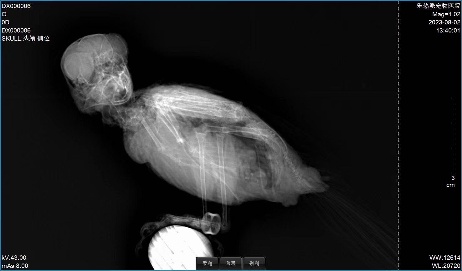
Clinical Imaging Samples
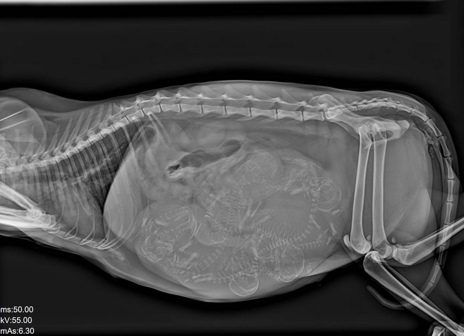
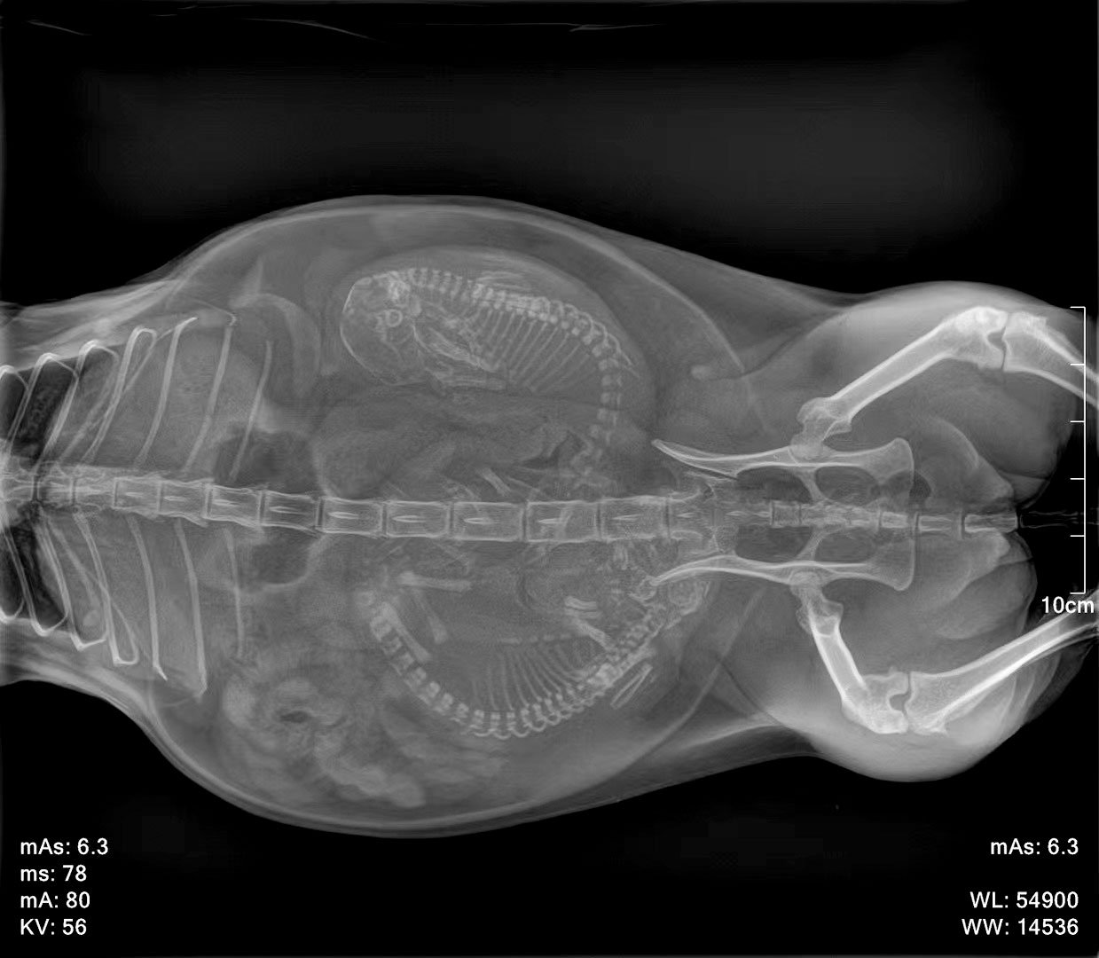
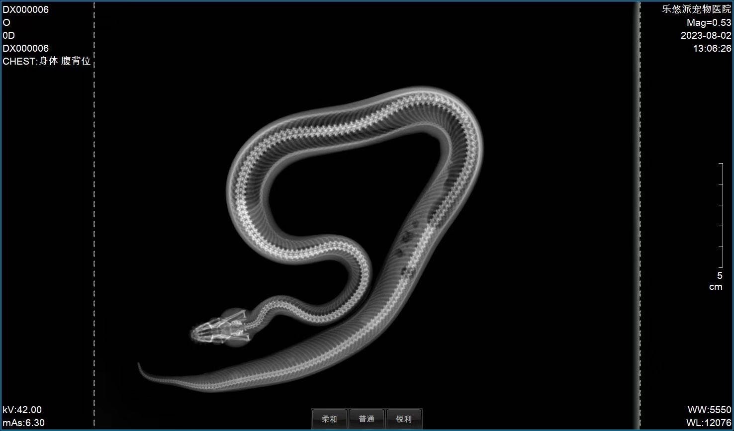
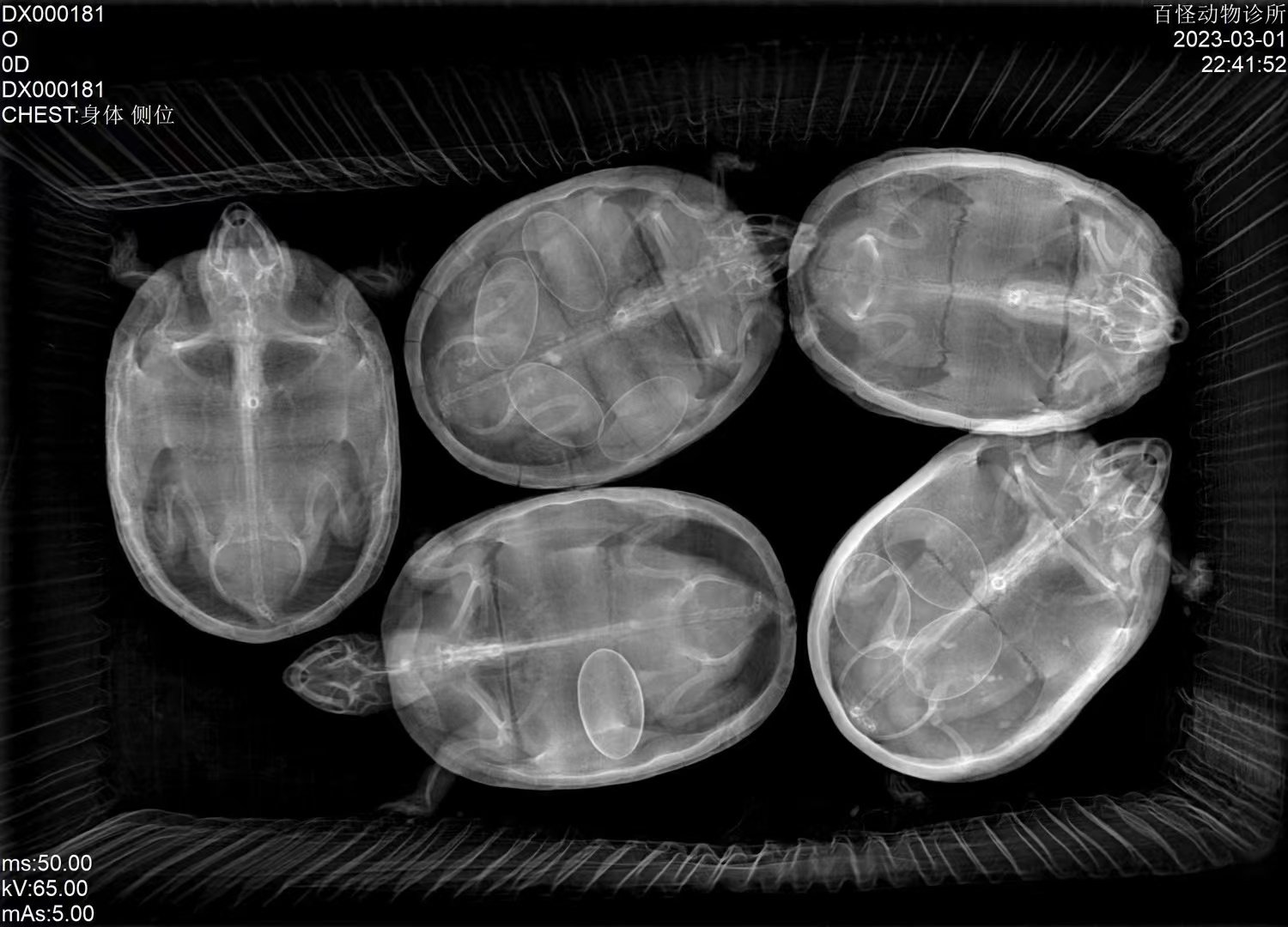
Representative radiographs captured on iPet-400 across companion-animal and exotic species, illustrating image quality across body sizes and tissue contrast.
Workflow & Installation
Single-phase 110/220 VAC power, no dedicated three-phase supply required. Installation footprint accommodates the 1.4 m patient table with overhead tube column. Operating environment: 5–35 °C, 10–75 % RH. Engineer-led installation and on-site operator training are included.
iPet-400 installation and operator training walkthrough (English narration, 6 min)
Package Contents
- High-voltage generator + control box
- X-ray tube assembly (Canon E7239X) + high-voltage cables
- 17×17″ CsI flat-panel detector + tethered power / data adapter
- LED collimator
- Patient table (1400×700 mm) with four-way floating top
- iVetXray workstation: 23.8″ display, 21.5″ touchscreen, PC, software dongle
- V-shape positioning set (1 large + 1 small)
- Lead protection set: gown, hat, scarf, glasses, gloves
- Foot switch, remote control, network cable (10 m)
Downloads
Why Choose IWA-DR01
- Workflow efficiency — 1-second acquisition with one-touch APR presets keeps room turnover high.
- Clinical accuracy — 16-bit CsI detector and Canon tube deliver consistent exposure for diagnostic-quality images.
- Practice economics — entry-level configuration with proven core components keeps installation and service cost predictable.
Warranty & Support
Two-year warranty covering hardware and software. Extended-warranty service is available; spare-parts price list is published for predictable maintenance budgeting. Engineer-led on-site installation and operator training included.
FAQ
Q: Does the system require three-phase power?
No. IWA-DR01 runs on single-phase 110/220 VAC.
Q: Is fluoroscopy supported?
No. IWA-DR01 is a static DR system. For dynamic fluoroscopy, see IWA-DR04 (iSmart-32) or IWA-DR05 (iSmart-32 Pro).
Q: Is the detector wireless?
No. The standard 17×17″ detector is tethered. A wireless detector is available with the IWA-DR02 (iPet-400 Pro) configuration.
Q: What PACS / DICOM standards are supported?
The workstation is DICOM 3.0 compliant for Store, Print, and Worklist with most third-party PACS.
Q: What patient size is supported?
The 1400×700 mm table top accommodates canine, feline, and exotic small-animal patients up to typical large-breed dogs.

Reviews
There are no reviews yet.