
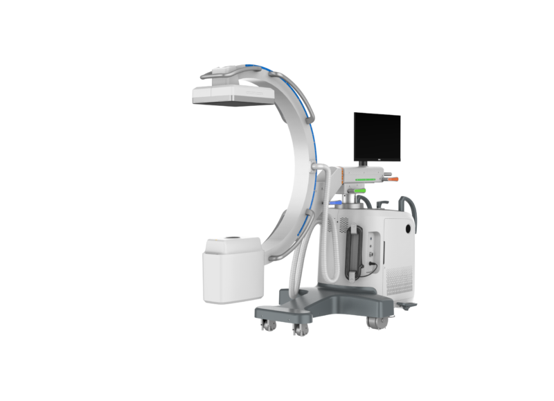
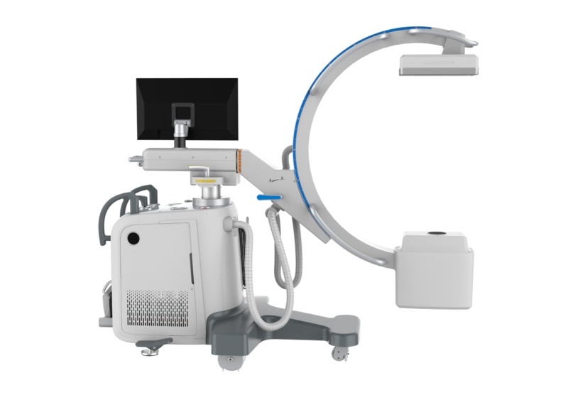
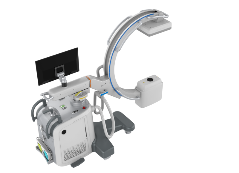
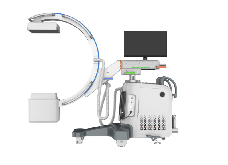
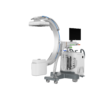
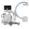
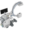
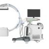
Mobile digital C-arm fluoroscopy system for veterinary orthopedic surgery, interventional procedures, and diagnostic imaging.
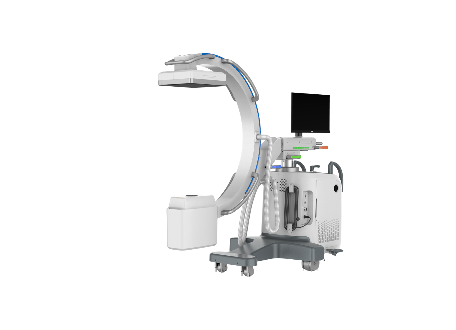
Product Overview
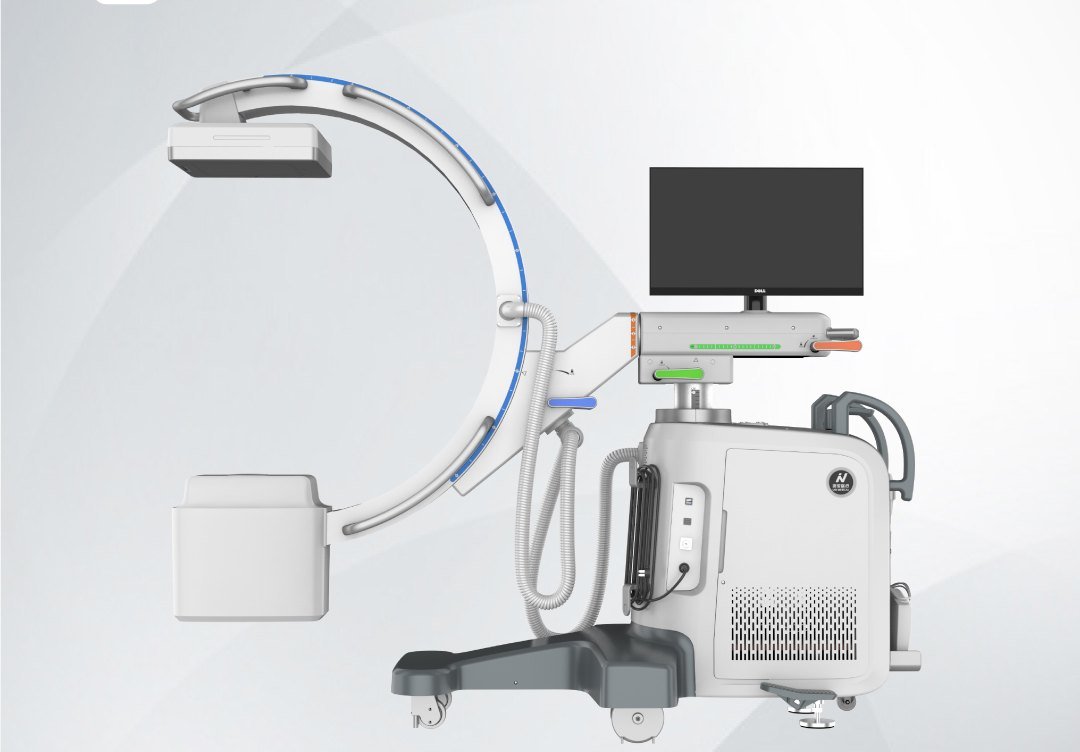
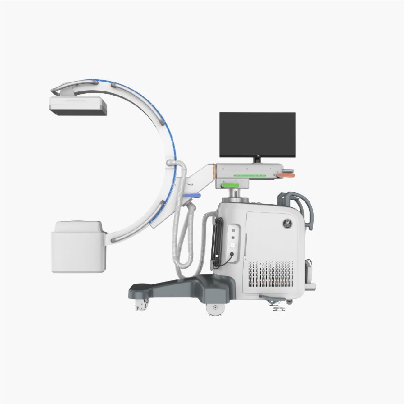
The IWAMED IWA-DC01 (iPet-C5) is a mobile digital C-arm fluoroscopy system built for small-animal and equine practices that perform orthopedic surgery, soft-tissue intervention, and diagnostic fluoroscopy. A 5 kW high-frequency generator drives a 40–125 kV tube paired with a 209.9 × 209.9 mm CsI flat-panel detector, delivering continuous and pulsed fluoroscopy plus digital spot and photographic acquisition from a single platform.
The free-sliding C-arm reaches the imaging field with multi-axis motion — orbital slide, ±180° rotation, vertical and lateral translation, and ±15° lateral swing — so the surgeon repositions the gantry, not the patient. A touch-screen workstation drives acquisition, image processing and DICOM-ready review.
Key Features
- 5 kW high-frequency generator with 40–125 kV continuous adjustment in 1 kV steps for both small and large patients.
- Mercu0909F CsI flat-panel detector (209.9 × 209.9 mm, 205 µm pixel pitch, 16-bit / 65 536 grayscale) for low-dose, high-contrast imaging.
- Three live modes — continuous fluoroscopy (0.3–6.3 mA), pulsed fluoroscopy (0.3–32 mA) and digital spot (up to 100 mA) — selected per procedure to balance dose and motion freezing.
- Free-sliding C-arm with ±180° rotation, 150° orbital slide (-33°/+117°), 400 mm vertical, 200 mm lateral translation and ±15° lateral swing.
- 980 mm SID with 640 mm arc depth and 780 mm aperture clearance — accommodates equine limbs and large-breed canine patients on the surgical table.
- Touch-screen control with foot-switch exposure, removable anti-scatter grid and integrated dose-area-product (DAP) and air-kerma indication for radiation accountability.
- Crux603i electric collimator with CAN-bus control, ≤0.5 mGy/h leakage radiation (IEC 60601-1-3) and motorized field shaping.
- Compact 1886 × 1563 × 853 mm footprint on a single mobile chassis — moves between OR, fluoroscopy suite and recovery without re-installation.
Product Appearance
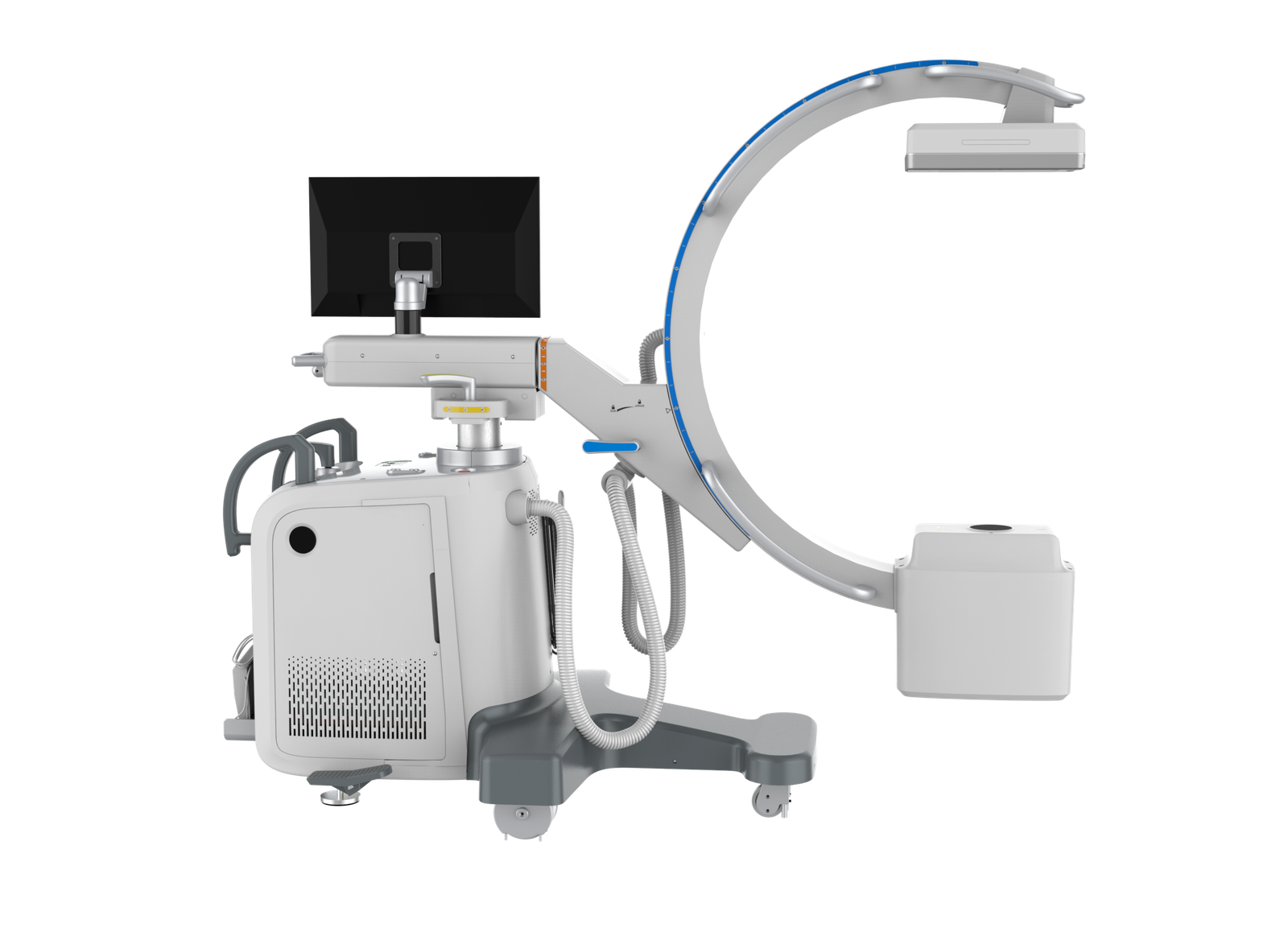
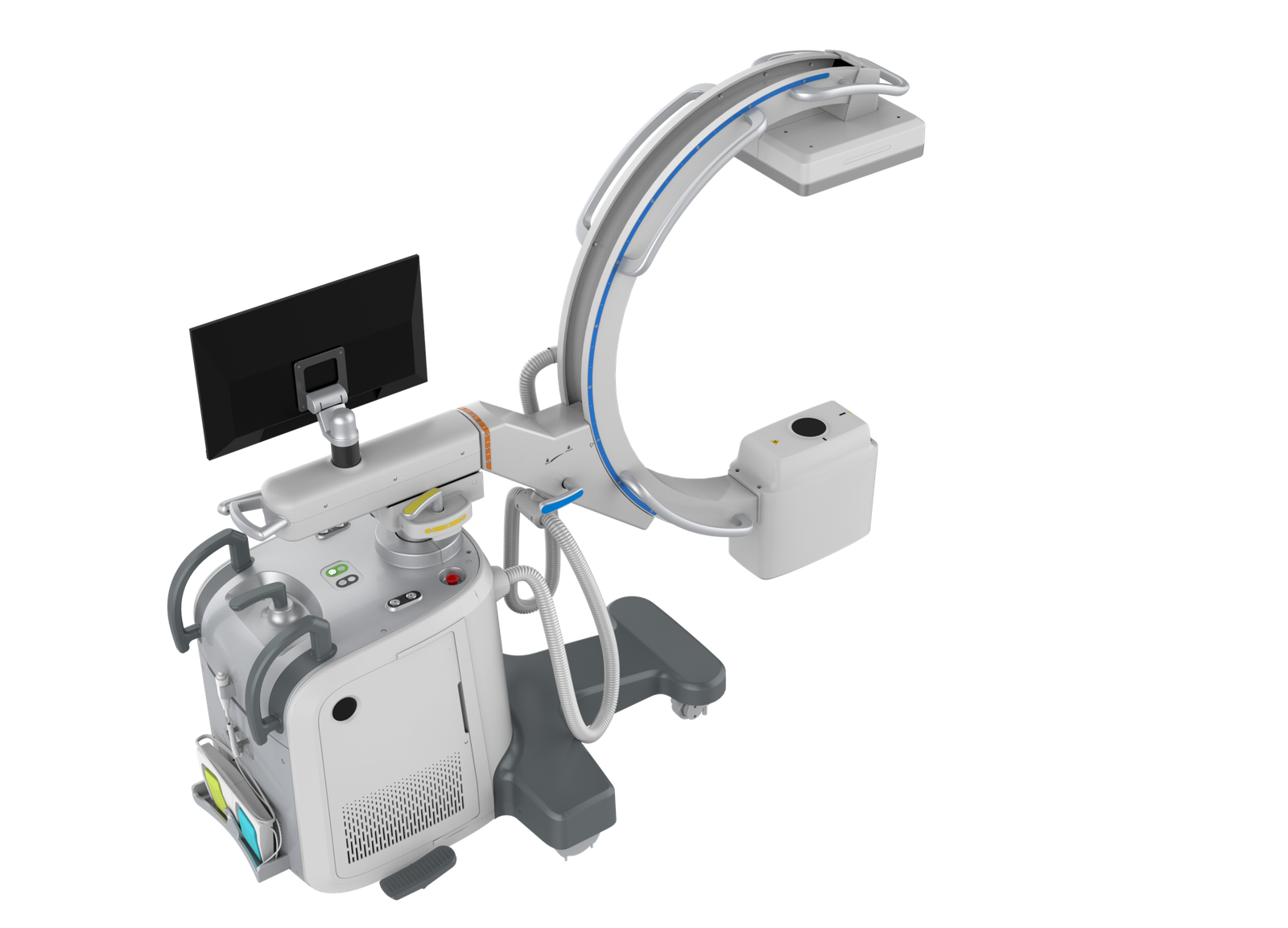
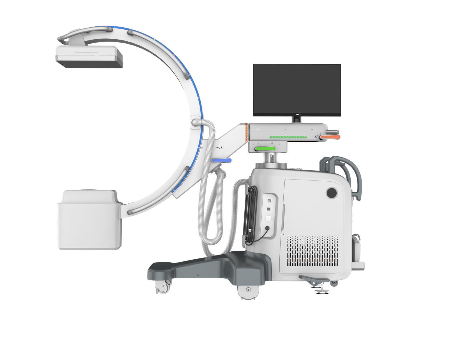
In the Operating Room
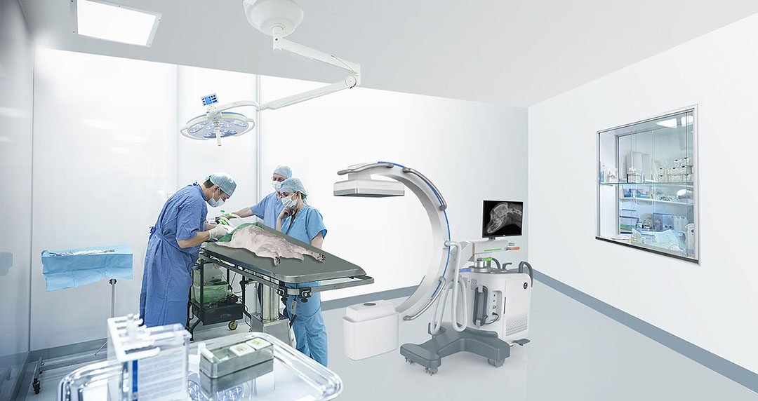
The C-arm is positioned beside the surgical table; surgeons work continuously while the detector arm provides intra-operative fluoroscopic guidance on a live monitor without breaking sterile field.
C-Arm Movement Capabilities
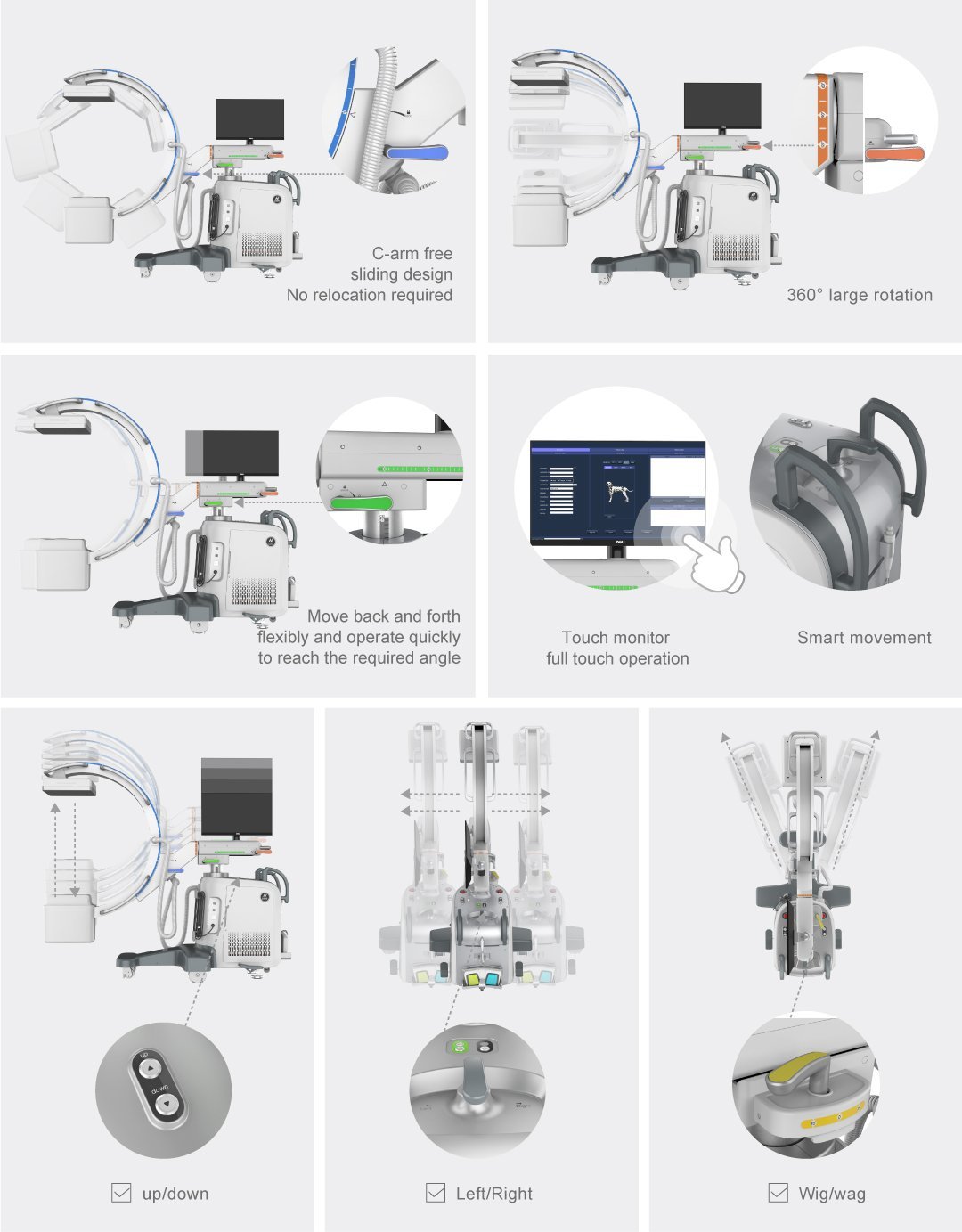
Eight independent motion axes let the operator bring the imaging field to the patient — never the reverse:
- C-arm free sliding design — orbital slide reaches different projections without relocating the chassis.
- 360° large rotation — full ±180° gantry rotation for orthogonal and oblique views.
- Forward / backward translation — horizontal travel reaches the surgical field with minimal repositioning.
- Touch monitor with full-touch operation — gantry-side touch display drives acquisition without keyboard or mouse.
- Smart movement — assist handles on the gantry head guide one-handed repositioning.
- Up / down vertical lift — vertical travel adjusts SID and operator working height.
- Left / right lateral translation — lateral travel covers wide surgical tables.
- Wig / wag (lateral swing) — fine ±15° lateral swing aligns the beam to oblique anatomy.
Operating Workflow
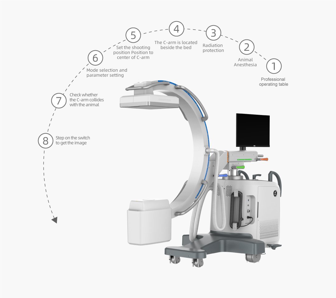
- Professional operating table — prepare a radiolucent surgical table compatible with C-arm clearance.
- Animal anaesthesia — induct and maintain the patient with standard veterinary anaesthesia protocol.
- Radiation protection — fit lead aprons, thyroid shields and gloves; clear unprotected personnel.
- C-arm positioned beside the bed — wheel the chassis into place; engage parking brakes.
- Set shooting position — orbit / rotate the C-arm to centre the anatomy in the field.
- Mode selection and parameter setting — pick continuous / pulsed fluoroscopy or digital spot; adjust kV / mA.
- Check for C-arm collisions — verify clearance around the animal, drape and surgical instruments.
- Step on the foot switch — expose; live image appears on the gantry and workstation monitors.
Technical Specifications
X-Ray Generator (Gemini5)
| Tube voltage range | 40–125 kV (1 kV step) |
| kV accuracy | ± 5 % |
| Tube current range | 0.1–100 mA |
| Nominal focal spot | 0.3 / 0.6 mm (IEC 336) |
| Anode target angle | 10° |
| Anode speed | 2700–3000 rpm |
| Inherent filtration | 0.7 mm Al eq. (IEC 522) |
| Additional filtration | 2.0 mm Al |
| Maximum anode heat capacity | 200 kHU |
| Maximum anode heat dissipation | 300 W |
| HV box heat capacity | 1200 kHU (900 kJ) |
| Nominal output power | 5 kW (100 kV, 50 mA, 0.1 s) |
Flat-Panel Detector (Mercu0909F)
| Scintillator | CsI (cesium iodide) |
| Effective imaging area | 209.9 × 209.9 mm |
| Pixel pitch | 205 µm |
| AD converter | 16-bit |
| Grayscale | 65 536 levels |
| Spatial resolution | ≥ 2.2 lp/mm |
| Image uniformity | < 2.2 % |
| Data interface | Gigabit Ethernet |
| Removable anti-scatter grid | Yes |
| Detector dimensions / weight | 270 × 275 × 54 mm / 5.5 kg ± 120 g |
Imaging Modes & Performance
| Continuous fluoroscopy | 40–110 kV · 0.3–6.3 mA · 0.1 mA step |
| Pulsed fluoroscopy | 40–125 kV · 0.3–32 mA · adjustable frame rate |
| Digital spot | up to 100 mA |
| Photography mode | 40–125 kV · up to 5 s exposure |
| mAs range | 0.1–100 mAs (R’10 series) |
| Exposure time | 10–1600 ms |
| Low-contrast resolution | ≤ 3 % (fluoroscopy and photography) |
| Dynamic range | ≥ 14 wedges fluoroscopy · ≥ 16 wedges photography |
| Fluoroscopy stabilisation | ≤ 2 s |
| Fluoroscopy recovery time | ≤ 4 min |
Collimator (Crux603i)
| Maximum X-ray field | 31 × 31 cm at 100 cm SID |
| Field error | ≤ 1 % of SID |
| Leakage radiation | < 0.5 mGy/h (IEC 60601-1-3) |
| Power supply | 24 V DC/AC (±10 %), max. 4 A |
| Control / communication | Electric · CAN bus |
| Dimensions / weight | 249 × 122 × 106 mm / 3.7 kg ± 300 g |
C-Arm Mechanical Range
| Source-to-image distance (SID) | 980 mm ± 10 mm |
| Arc depth | 640 mm ± 10 mm |
| Aperture (opening) | 780 mm ± 10 mm |
| Arc radius | 520 mm ± 5 mm |
| Orbital slide | 150° (-33° / +117°) |
| Rotation | ± 180° |
| Vertical movement | 400 mm ± 8 mm |
| Lateral (horizontal) movement | 200 mm ± 4 mm |
| Lateral swing | ± 15° |
Workstation
| CPU | Intel Core i5 |
| Memory | 16 GB |
| Storage | 1 TB HDD |
| Operating system | Windows 10 64-bit |
| Gantry monitor | Dell P2418HT · 1920 × 1080 |
| Software | X-ray acquisition, processing & display |
Power, Environment & Footprint
| Power supply | Single-phase 220 V AC, 50 Hz ± 1 Hz |
| Voltage tolerance | ± 10 % of nominal |
| Power capacity | 9 kVA |
| Power resistance | ≤ 1 Ω |
| Operating temperature | 15–35 °C |
| Operating humidity | 35–75 % RH |
| Main frame dimensions | 1886 × 1563 × 853 mm |
| Weight | ≈ 280 kg |
| Shipping crate (L × W × H) | 220 × 160 × 155 cm · ≈ 300 kg |
Clinical Applications
Workflow & Installation Notes
The IWA-DC01 ships as a single mobile chassis and is designed to operate in a dedicated, lead-shielded fluoroscopy or surgical suite. Site requirements are documented in the iPet-C5 installation drawing supplied with the system.
- Power: single-phase 220 V AC ± 10 %, 50 Hz ± 1 Hz, 9 kVA capacity. The system requires its own dedicated supply with isolated neutral and earth — do not share with other equipment.
- Room geometry: minimum ceiling height 2300 mm; door width ≥ 950 mm; floor levelness within 1.5 mm and concrete substrate > 100 mm.
- Radiation shielding: ≥ 3 mm Pb equivalent in the primary beam direction, 2 mm Pb equivalent on remaining surfaces. Engage a qualified radiation-protection contractor and follow local regulations.
- Environment: 15–35 °C, 35–75 % RH; air-conditioning and dehumidification are recommended. Maintain forced negative-pressure ventilation in the imaging room.
- Cable conduit: 80 mm diameter floor-level pass-through between control and exam rooms.
- Workstation desk and operator console: supplied by the practice; mount the gantry-side touch monitor on the C-arm chassis.
Final layout, shielding calculation and acceptance testing are the responsibility of the local radiation-protection authority and the installing distributor.
Package Contents
- 1 × C-arm gantry with integrated X-ray tube housing (Gemini5 generator)
- 1 × Mercu0909F CsI flat-panel detector with removable anti-scatter grid
- 1 × Crux603i electric collimator
- 1 × Mobile chassis with high-voltage box and inverter assembly
- 1 × Workstation host (Intel Core i5, 16 GB RAM, 1 TB HDD, Windows 10) with X-ray acquisition software
- 1 × Touch-screen monitor (Dell P2418HT, 1920 × 1080) on gantry mount
- 1 × Foot-switch exposure controller
- 1 × Power cable set and operator manual
Downloads & Documentation
Why Choose the IWA-DC01
- Procedure throughput. Free-sliding C-arm reaches the imaging field in seconds — the surgeon does not move the patient or break sterile field to reposition for orthogonal views.
- Imaging confidence. 209.9 × 209.9 mm CsI flat panel at ≥ 2.2 lp/mm and 16-bit grayscale produces consistent low-contrast resolution (≤ 3 %) across fluoroscopy and photographic modes.
- Dose accountability. Continuous and pulsed fluoroscopy modes, real-time air-kerma indication and cumulative DAP reporting support ALARA workflows and radiation-safety auditing.
- Service economics. Standard Windows 10 workstation, gigabit Ethernet detector interface and CAN-bus collimator simplify field service, software updates and parts replacement.
Warranty & Support
IWAMED supplies the IWA-DC01 with a manufacturer warranty, installation supervision and operator training delivered through the local distributor. Coverage period and on-site service terms vary by territory — contact us for a written quotation specific to your jurisdiction.
FAQ
- What animal sizes does the IWA-DC01 accommodate?
- The 780 mm C-arm aperture and 640 mm arc depth cover canine and feline patients on a standard surgery table and accept equine distal limbs in standing or recumbent positioning. Patient mass is limited only by the surgical table itself, not the gantry.
- Does the system support DICOM export?
- The workstation runs general X-ray acquisition and display software with gigabit Ethernet to the detector. DICOM Store/Worklist integration with practice PIMS is available — confirm the exact DICOM service classes with your distributor at order time.
- What dose-management features are included?
- Continuous and pulsed fluoroscopy modes (with adjustable frame rate), removable anti-scatter grid, air-kerma rate display in mGy/min and cumulative air-kerma plus dose-area-product readouts visible from the operator position.
- What site preparation is required before delivery?
- A dedicated 220 V / 9 kVA supply, ≥ 2300 mm ceiling height, ≥ 950 mm door width, lead-shielded room walls (3 mm Pb primary, 2 mm Pb secondary), 80 mm floor cable conduit and a controlled environment of 15–35 °C / 35–75 % RH. Shielding design must be reviewed by your local radiation-protection authority.
- Is the C-arm fixed or mobile?
- The IWA-DC01 is a mobile chassis on castors. It moves between operating room, imaging suite and emergency area without re-installation. Daily relocation does not require recalibration when the floor surface meets the 1.5 mm levelness specification.
Reviews
There are no reviews yet.