
Micro-scale CT for exotic patients — a self-shielded, mobile CBCT with a 50 µm focal spot and 0.05 mm slices.
Product Overview
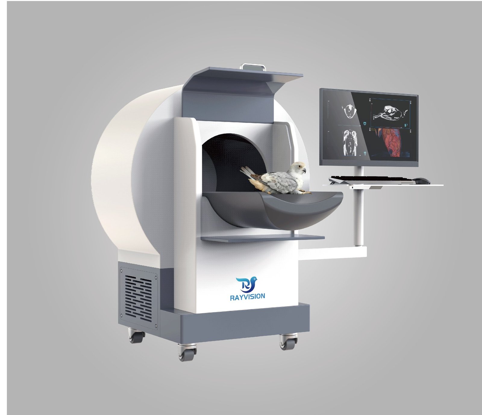
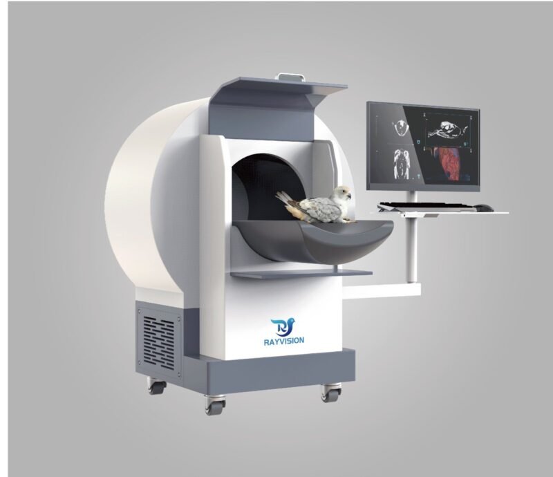
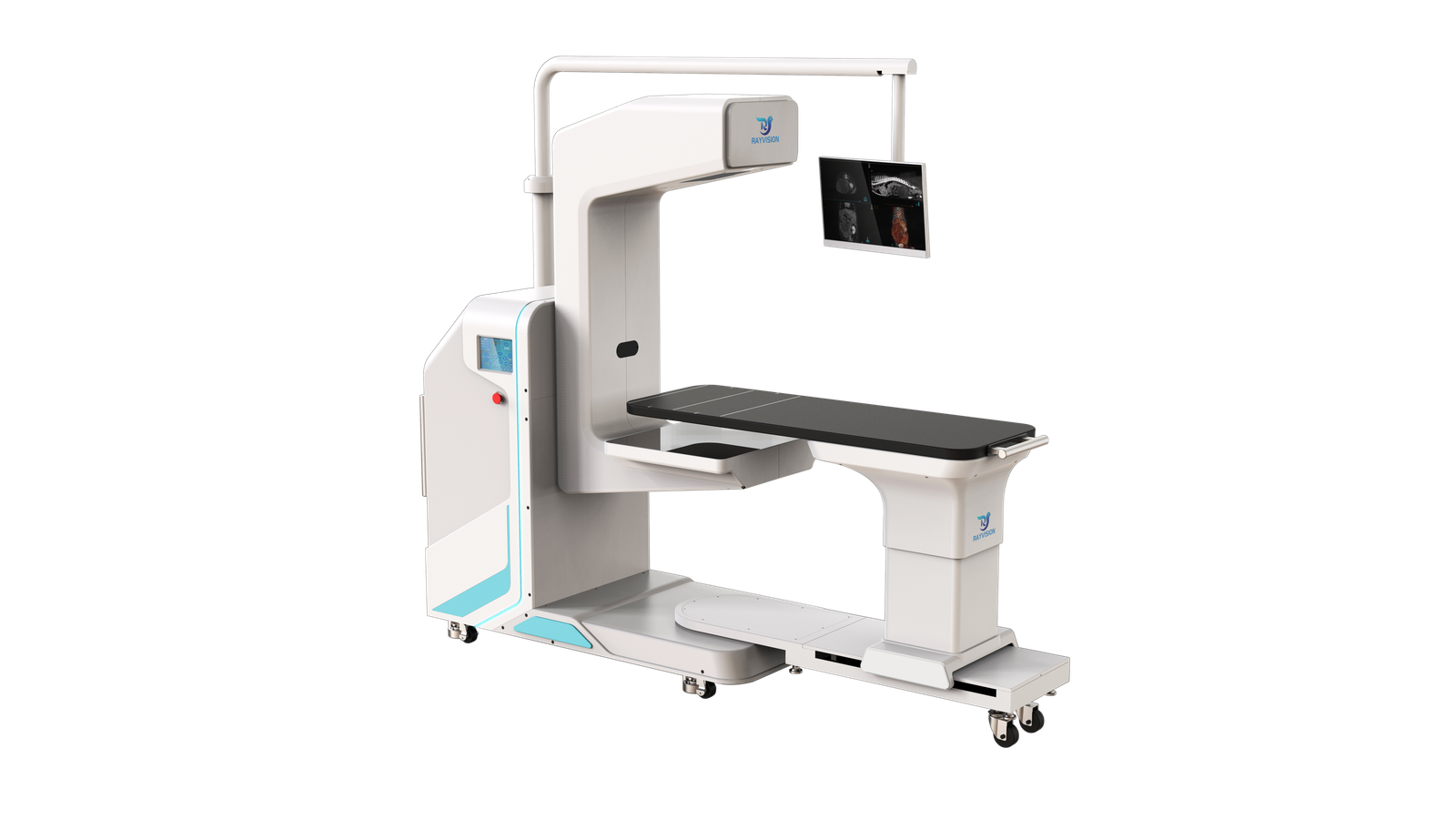
Micro50 is a mobile veterinary cone-beam CT system from Rayvision Medical, dedicated to exotic species. A 50 µm focal spot and slice thickness down to 0.05 mm resolve fine structures in small exotic patients and in juvenile feline and canine patients.
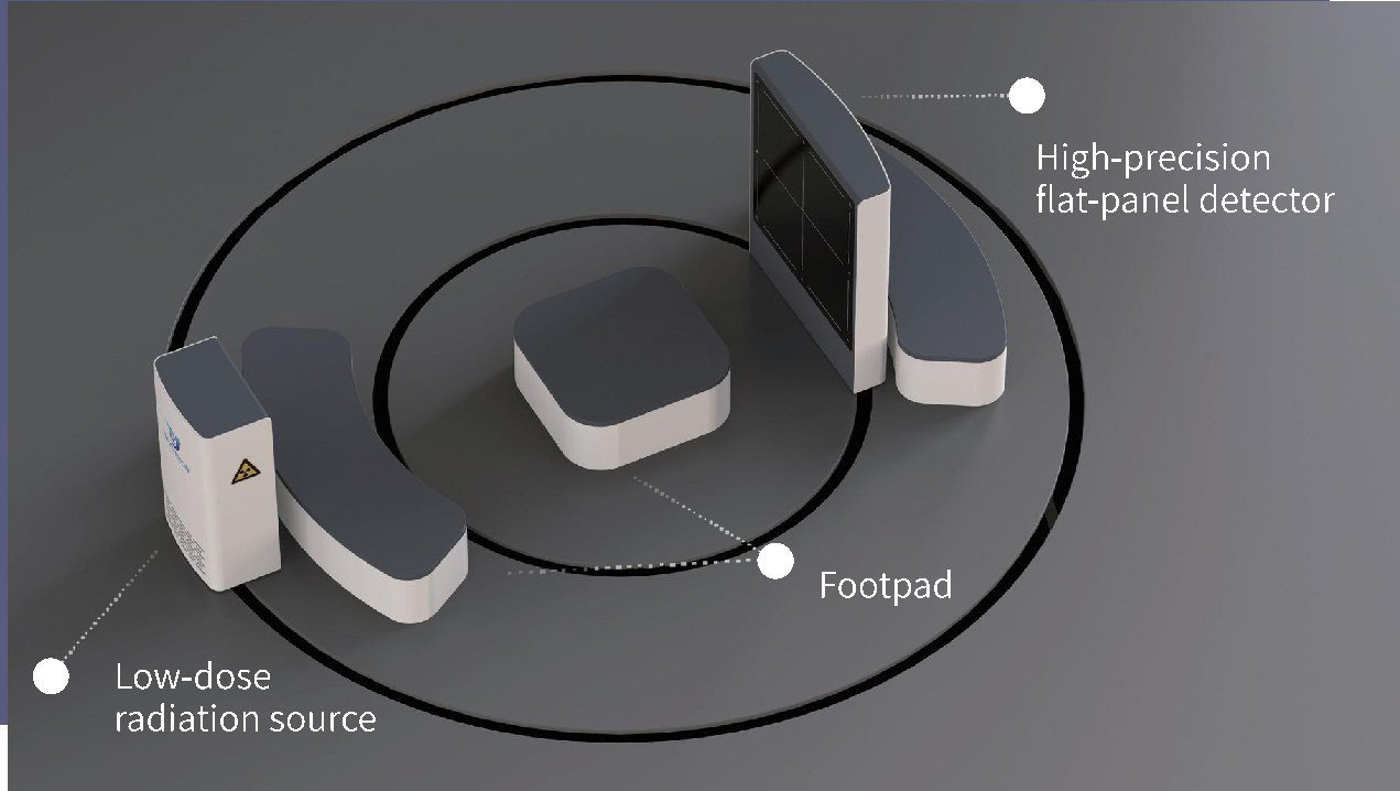
The self-shielding enclosure removes the need for a dedicated shielded room (equipment surface radiation ≤ 2.5 µSv/h). The system runs on castors, needs a 1 kW supply, and moves between consulting rooms and operating theatres. A machine-side display lets the operator adjust positioning and review images at the gantry.
Key Features
- Self-shielding enclosure — no dedicated shielded room; equipment surface radiation ≤ 2.5 µSv/h.
- Micro-imaging — 50 µm focal spot, 0.05 – 0.24 mm slice thickness, 600 reconstructed slices, 140 µm detector pixel.
- 3-in-1 imaging — CT, fluoroscopy (real-time dynamic enhancement, 1 minute continuous exposure) and DR (edge enhancement, TPLO / VHS / TTA tools).
- Mobile and ready to use — castor-mounted; no complex installation.
- Machine-side display — position and review at the gantry without walking back and forth.
- Positioning — scan table travel 800 mm horizontal / 300 mm vertical, laser positioning light, 330 mm aperture.
Product Gallery
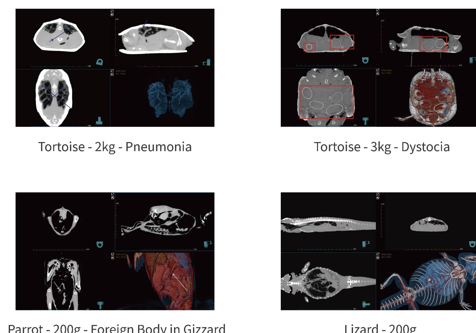
Clinical Imaging Samples
Exotic imaging cases acquired on Micro50.
Technical Specifications — X-ray Source
| Focal spot | 50 µm |
| Tube voltage | 30 – 80 kV |
| Tube current | 50 – 700 µA |
Technical Specifications — Power Supply
| Rated power | 1 kW |
| Input power | 220 VAC ±10 % |
Technical Specifications — Ergonomics
| Aperture | 330 mm |
| Scan table travel | Horizontal 800 mm / vertical 300 mm |
| Positioning | Laser positioning light |
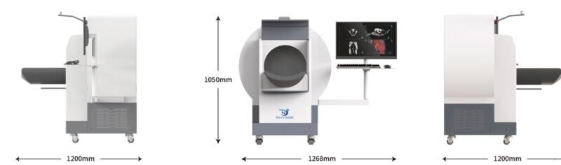
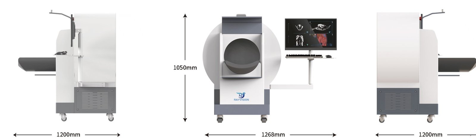
| Overall dimensions (drawing) | 1268 mm (W) × 1200 mm (D) × 1050 mm (H) |
Technical Specifications — Imaging Parameters
| Reconstructed slices | 600 |
| Slice thickness | 0.05 – 0.24 mm |
| Maximum scan diameter | [TBC] |
| Stitched scan length | [TBC] |
| Scan time | 15 – 60 s |
| Detector pixel | 140 µm |
| Maximum acquisition speed | 30 fps |
| Detector active area | 30 × 25 cm |
Clinical Applications
- Chelonians — respiratory disease (pneumonia) and reproductive disorders (dystocia).
- Avian patients — gastrointestinal foreign bodies, including ventricular (gizzard) foreign bodies in psittacines.
- Lizards and other small reptiles — whole-body CT of patients around 200 g.
- Juvenile feline and canine patients — high-detail imaging of small anatomy.
Site Planning and Installation
Micro50 is self-shielded and mobile. The manufacturer states that the shielding performance complies with applicable standards; confirm local radiation-licensing requirements before use.
Power and Environment
| Supply voltage | 220 VAC ±10 % |
| Supply internal resistance | ≤ 0.5 Ω |
| Frequency | 50 Hz / 60 Hz |
| Power capacity | 1 kW |
| Operating temperature | 10 °C – 35 °C |
| Relative humidity | 30 % – 75 %, non-condensing |
| Atmospheric pressure | 700 – 1060 hPa |
| Equipment surface radiation | ≤ 2.5 µSv/h |
Workflow Notes
- Wheel the system into a consulting room or operating theatre and connect it to a 220 VAC supply.
- Position the patient on the scan table with the laser light and confirm on the machine-side display.
- Select CT, fluoroscopy or DR acquisition; CT scans take 15 – 60 s.
Package Contents
- 1 × Micro50 gantry with integrated scan table and laser positioning light
- 1 × Machine-side display on articulated arm with keyboard tray
Downloads
Product brochures and technical documentation are available on request — contact IWA.
Why Choose Micro50
- Workflow efficiency — The system comes to the patient: mobile, 1 kW powered, with a machine-side display for positioning and review.
- Clinical accuracy — A 50 µm focal spot and 0.05 mm slices resolve structures that standard CT misses in patients weighing a few hundred grams.
- Practice economics — Self-shielding removes shielded-room construction and renovation costs.
Warranty and Support
1-year whole-system warranty. Extended warranty is available for purchase — contact IWA for terms and service coverage in your region.
Installation is carried out against the manufacturer’s installation manual: the main unit and patient table are connected, the console and workstation are cabled, and scanning, reconstruction, fluoroscopy and DR acquisition are verified before handover.
FAQ
Q: Does Micro50 need a shielded room?
No dedicated shielded room is required — the enclosure is self-shielding and equipment surface radiation is ≤ 2.5 µSv/h. Local radiation-licensing rules still apply.
Q: What size of patient fits?
The aperture is 330 mm. The system is intended for exotic species and small juvenile feline and canine patients; confirm larger patients with IWA.
Q: What power supply does it need?
220 VAC ±10 %, 50 Hz or 60 Hz, 1 kW, supply internal resistance ≤ 0.5 Ω.
Q: Can it perform radiography as well as CT?
Yes. Micro50 is 3-in-1: CT, fluoroscopy and DR with edge enhancement and TPLO / VHS / TTA tools.
Q: What warranty applies?
1-year whole-system warranty, with extended warranty available for purchase.
Reviews
There are no reviews yet.