
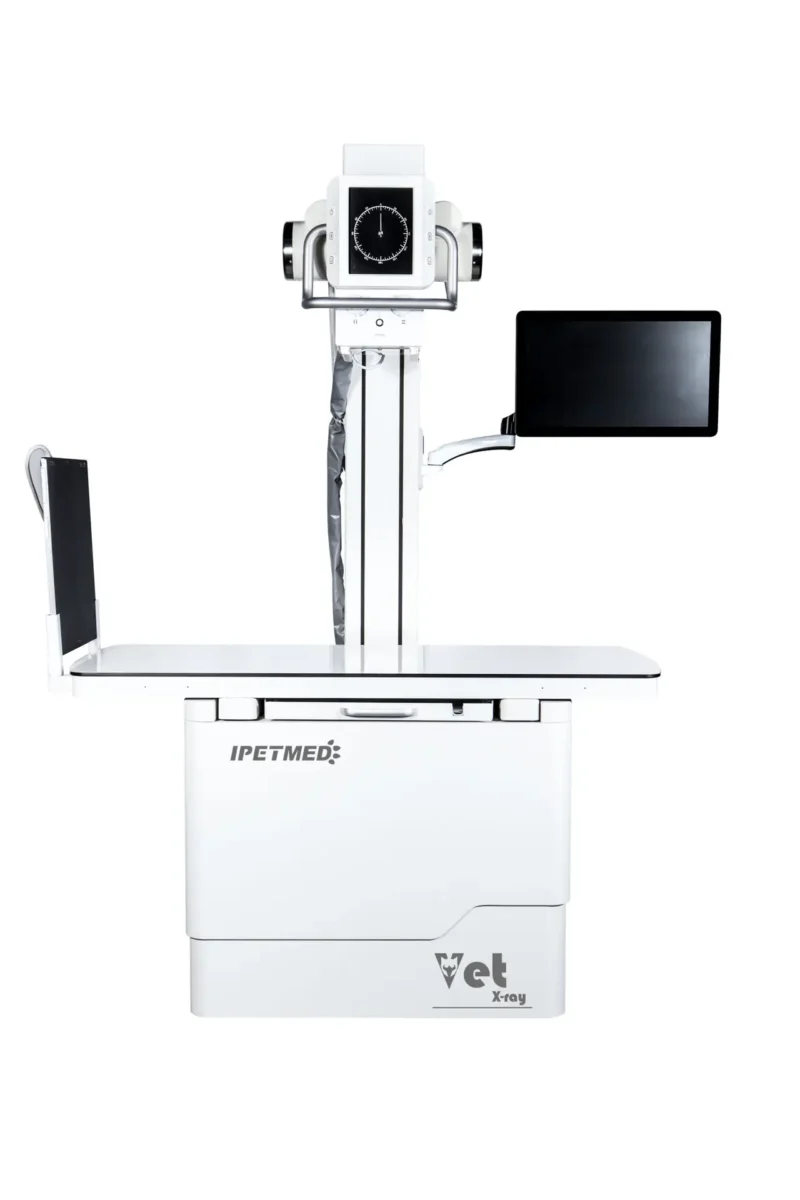
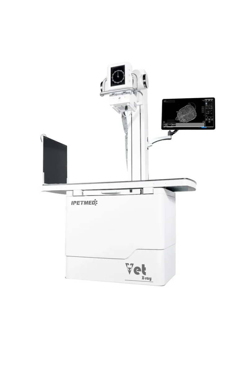
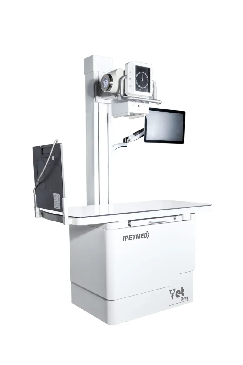
Upgraded multifunctional veterinary DR system with removable column and 0.6 / 1.2 mm dual-focus tube, ready for both tabletop and wall-stand workflows.
Product Overview
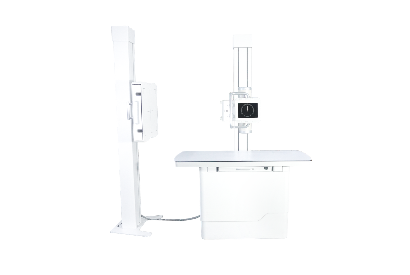
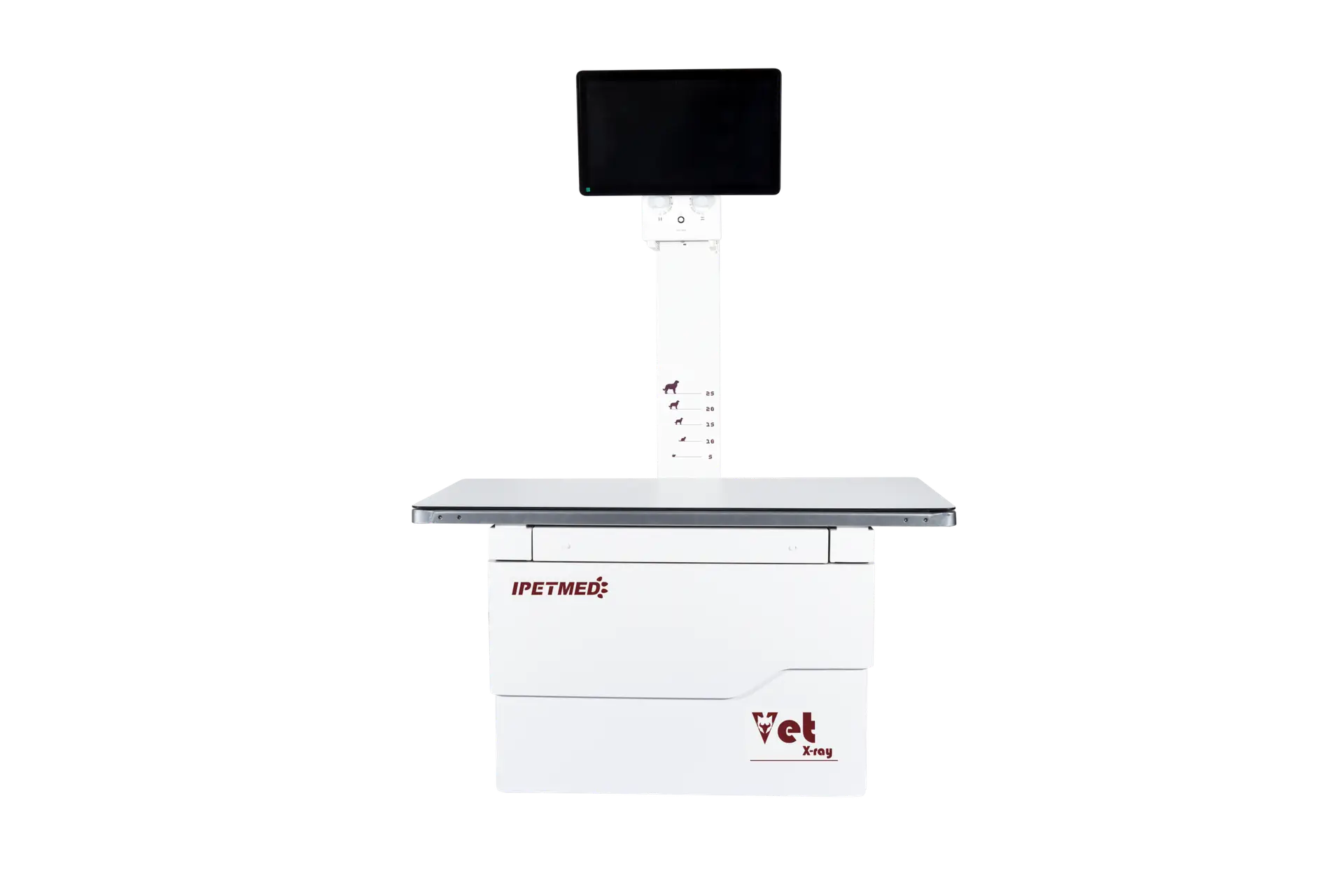
IWA-DR02 iPet-400 Pro Veterinary DR X-ray Imaging System extends the iPet-400 platform with finer focal-spot geometry and a removable column that turns a single installation into a tabletop-plus-vertical-bucky setup. The 32 kW high-frequency generator, Canon E7240X dual-focus tube (0.6 / 1.2 mm), and 17×17″ CsI flat-panel detector deliver one-second acquisition for daily small- and medium-animal practice.
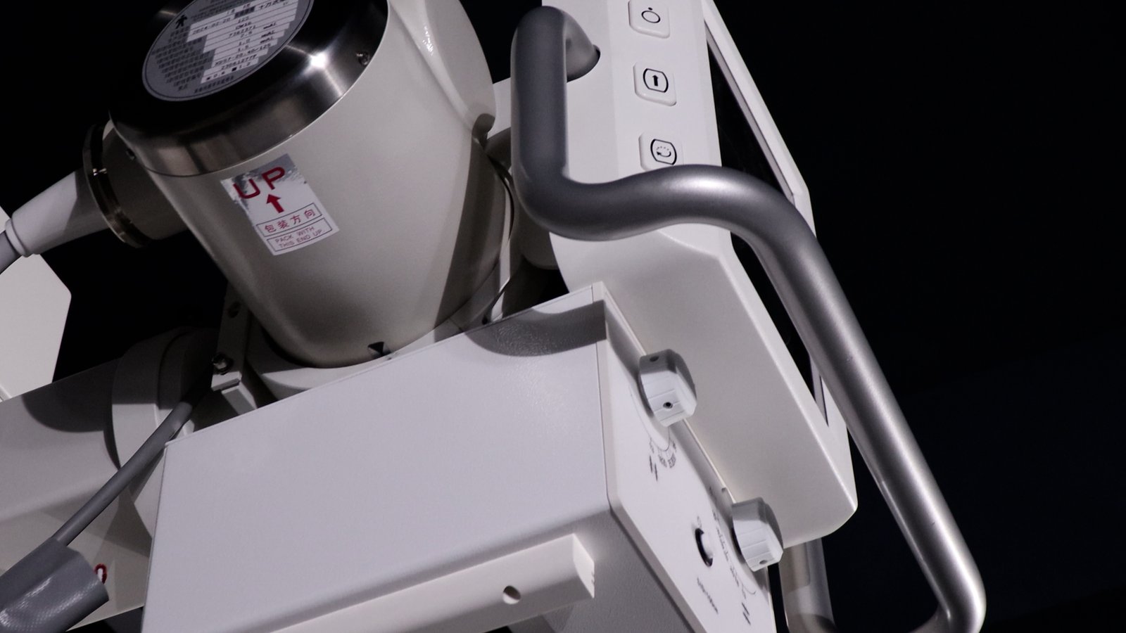
The removable column allows the X-ray tube to rotate and reposition for thorax shots in standing horses, large dogs, and any case where a vertical film is preferred to bench-top imaging.
Key Features
- Removable column — switch between tabletop radiography and vertical-stand imaging without changing rooms.
- 0.6 / 1.2 mm dual-focus Canon E7240X tube — fine detail for dental and small-bone work, large focus for high-throughput exposures.
- 32 kW high-frequency generator: 40–150 kV, 10–400 mA, 1–10000 ms exposure range.
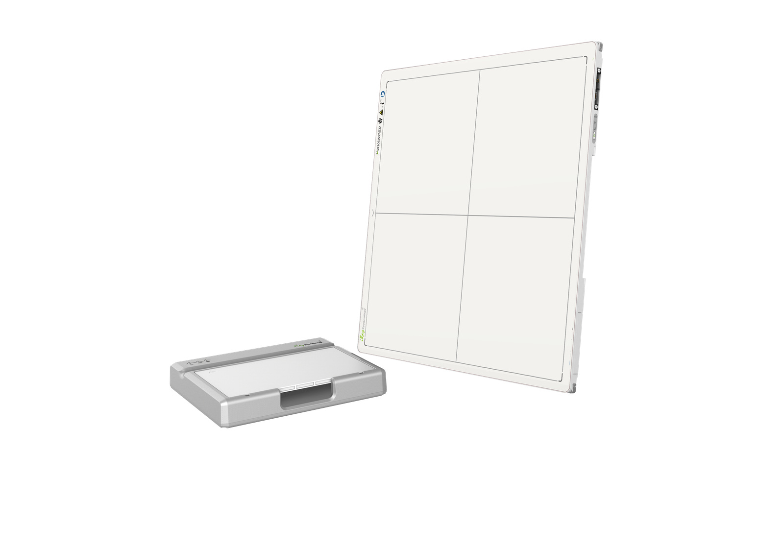
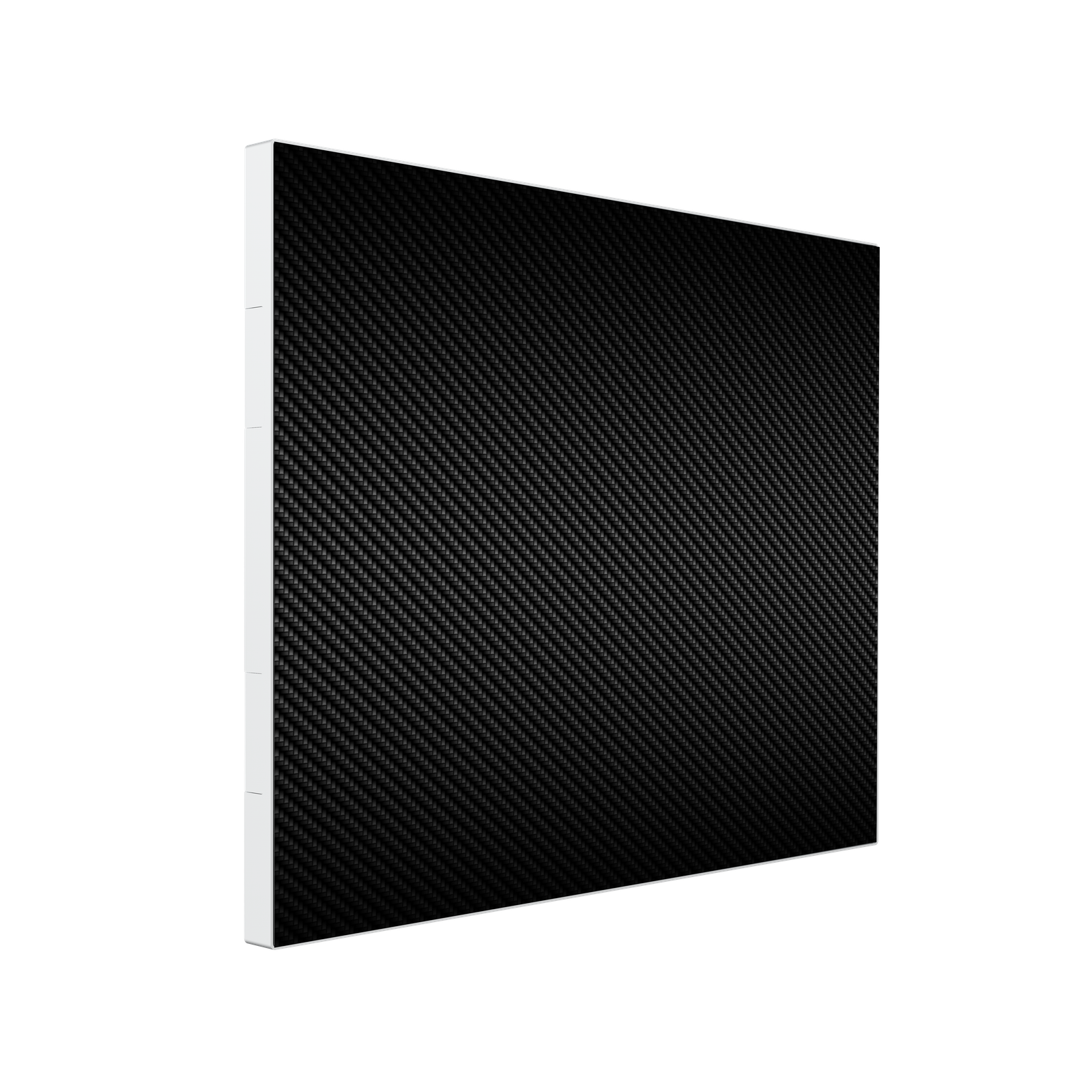
- 17×17″ CsI flat-panel detector: 3072×3072, 139 µm pitch, 16-bit A/D, 3.6 LP/mm, 1-second acquisition.
- Optional wireless detector for fully mobile workflow in surgical theatres and large-animal stalls.
- iVetXray + MiniPACS workstation with veterinary measurement toolkit (VHS, TPLO, HIP, TTA) and DICOM 3.0 PACS integration.
- Dual-display interface: 23.8″ diagnostic monitor + 21.5″ tableside touchscreen.
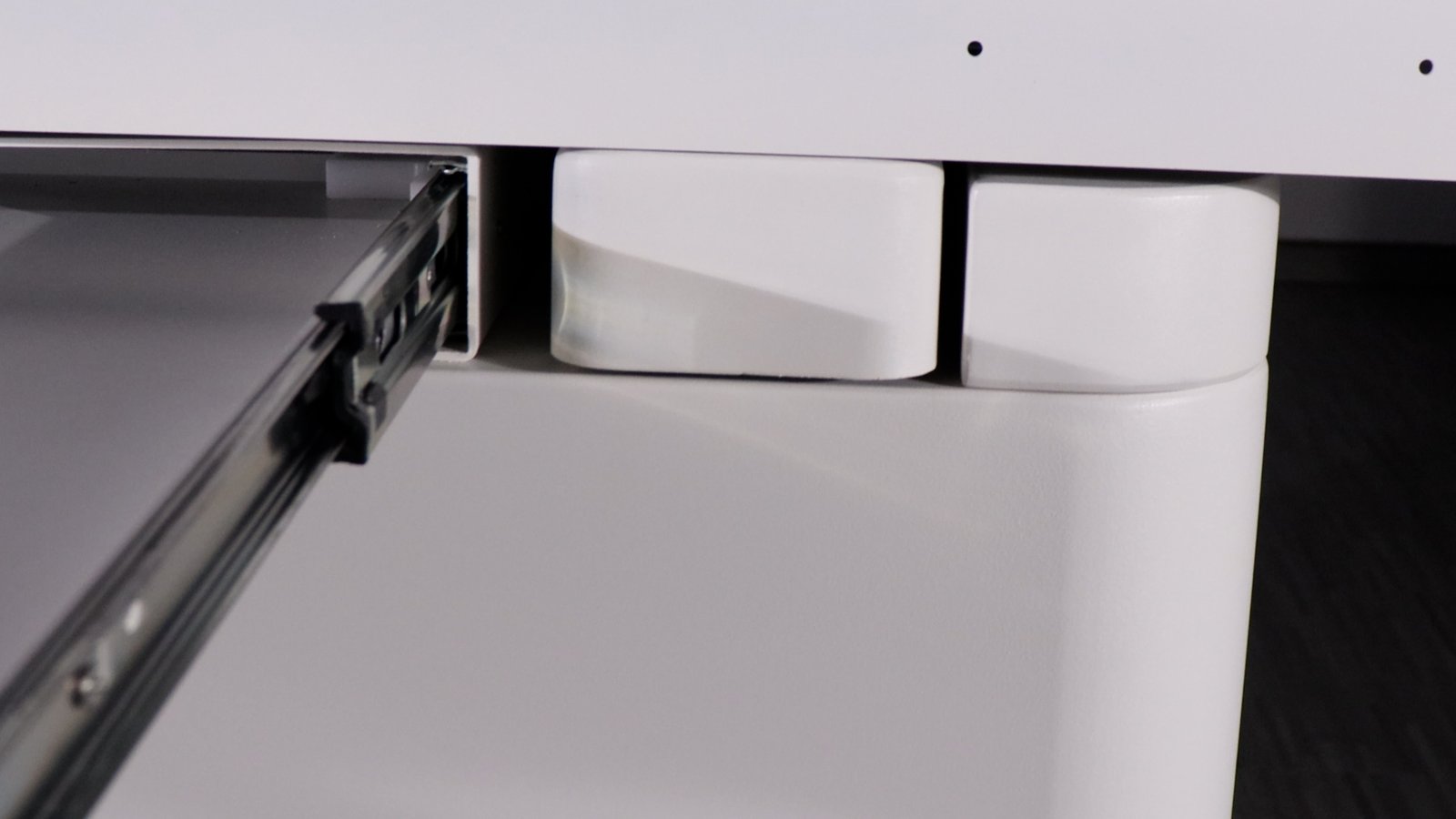
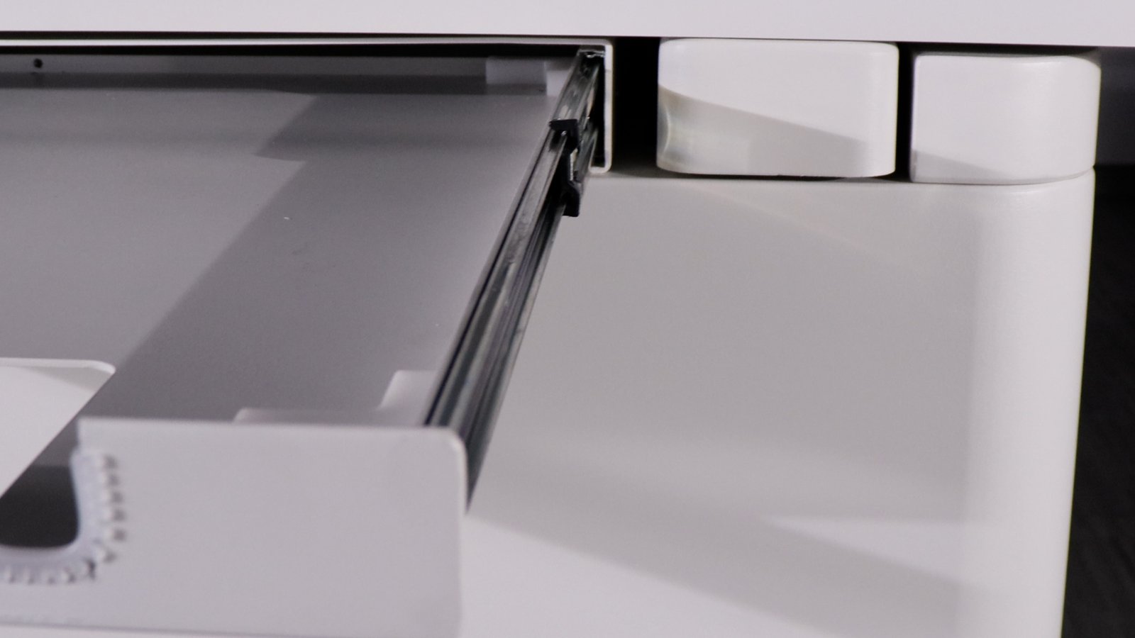
- Four-way floating table 1400×700 mm, phenolic resin top, ±75 mm longitudinal and ±200 mm lateral travel.
- Stable LED collimator with 1.0 mm Al filtration at 75 kV.
Product Gallery
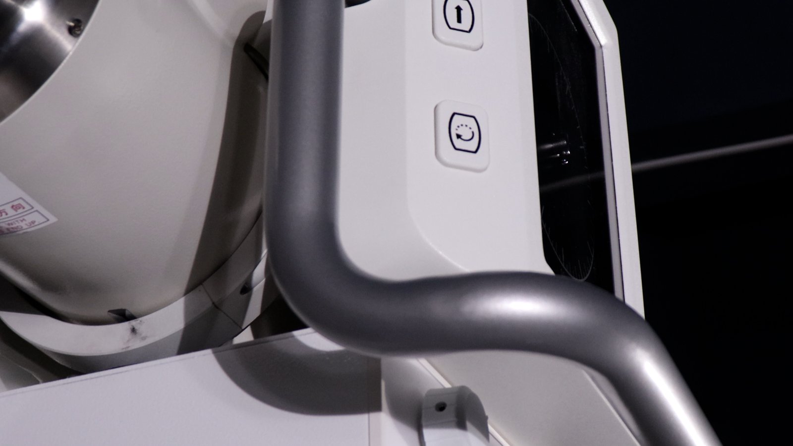
Detail views of the removable column, X-ray tube head, four-way floating patient table, LED collimator, and operator workstation.

High-voltage generator
| Output power | 32 kW |
| kV range | 40 – 150 kV |
| mA range | 10 – 400 mA |
| mAs range | 0.1 – 100 mAs |
| Exposure time | 1 – 10000 ms |
| Power input | Single phase, 110/220 VAC ±10% |
X-ray tube (Canon E7240X)
| Max voltage | 125 kV |
| Focal spot | 0.6 / 1.2 mm (dual) |
| Anode input power | 15 kW (small) / 30 kW (large) |
| Anode heat capacity | 140 kHU |
Flat-panel detector
| Technology | a-Si TFT, CsI scintillator |
| Image size | 43 × 43 cm (17×17″) |
| Pixel matrix | 3072 × 3072 |
| Pixel pitch | 139 µm |
| A/D conversion | 16 bit |
| Spatial resolution | 3.6 LP/mm |
| Image acquisition | 1 s |
| Data interface | GigE |
| Min. detectable dose (RQA5) | 14 nGy |
| Sealing | IP65 |
Column, table, collimator
| Column | Removable; SID adjustable; tube rotating for vertical film |
| Table size | 1400 × 700 mm, phenolic resin top |
| Table movement | X: 75 ±10 mm, Y: 200 ±10 mm |
| Collimator light source | LED |
| Filter | 1.0 mm Al at 75 kV |
Workstation
| Software | iVetXray + MiniPACS |
| Display | 23.8″ diagnostic monitor + 21.5″ tableside touchscreen |
| OS | Windows 10 |
| Storage | 256 GB SSD + 1 TB HDD |
Clinical Applications
- Canine and feline general radiography (thorax, abdomen, skeletal)
- Vertical bucky shots for standing large-breed dogs or large animals
- Orthopaedic workflows: TPLO, TTA, hip dysplasia (VHS)
- Dental and skull imaging with the 0.6 mm fine focus
- Pre-anaesthetic and post-operative imaging
Clinical Imaging Samples
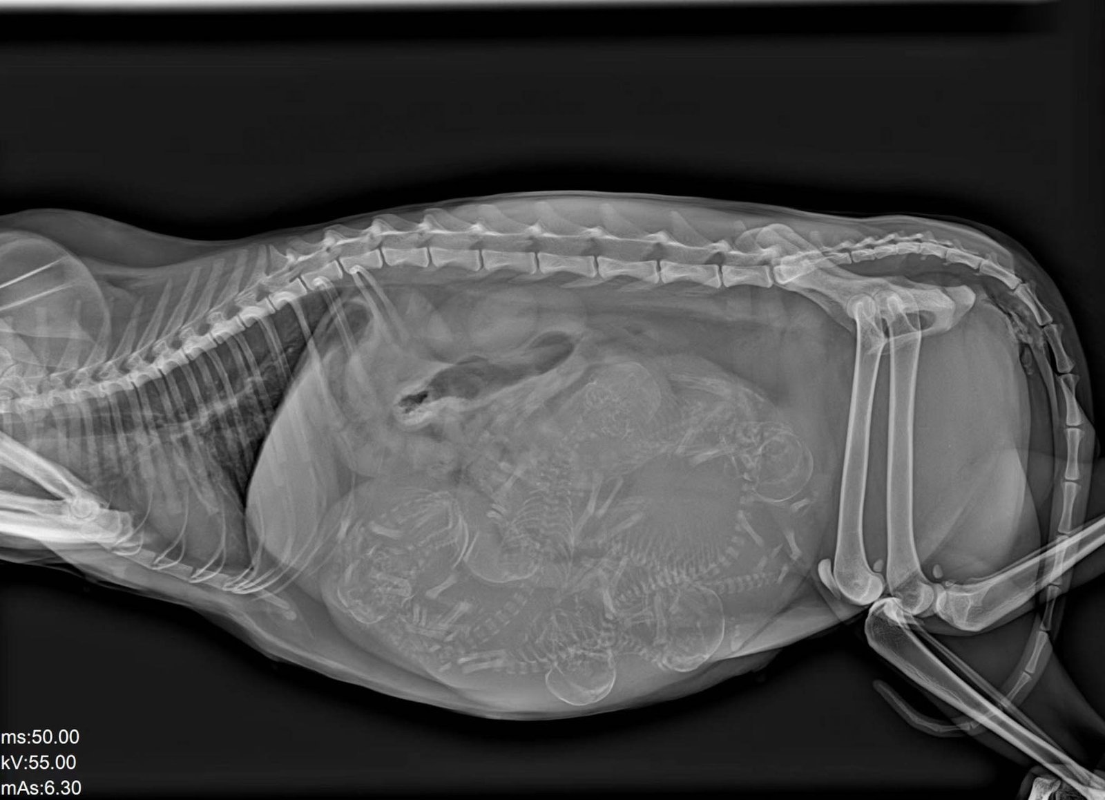
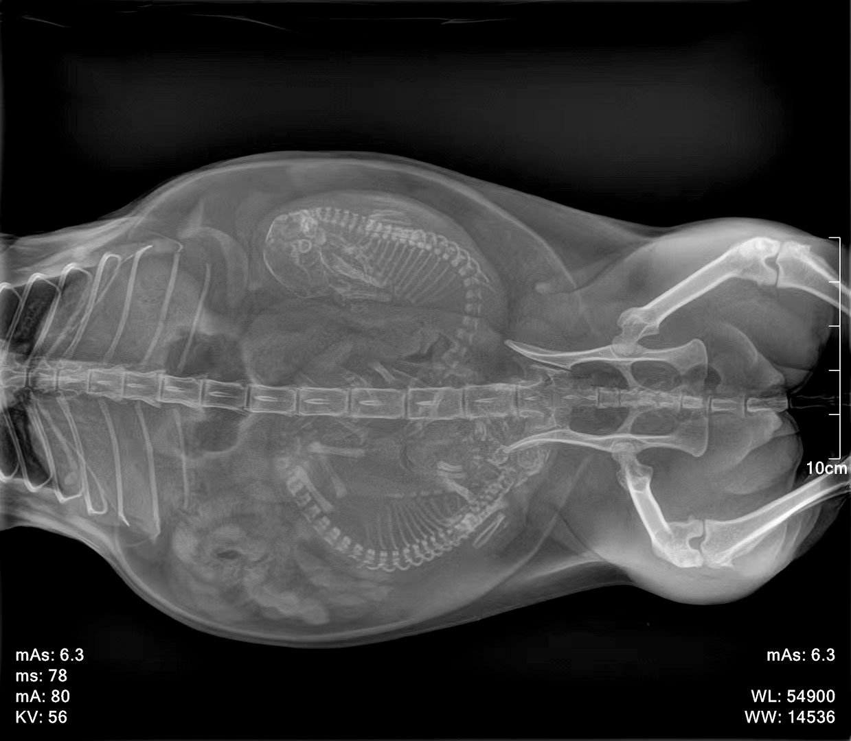
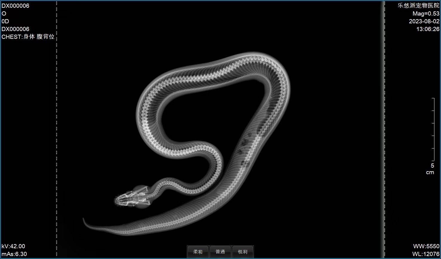
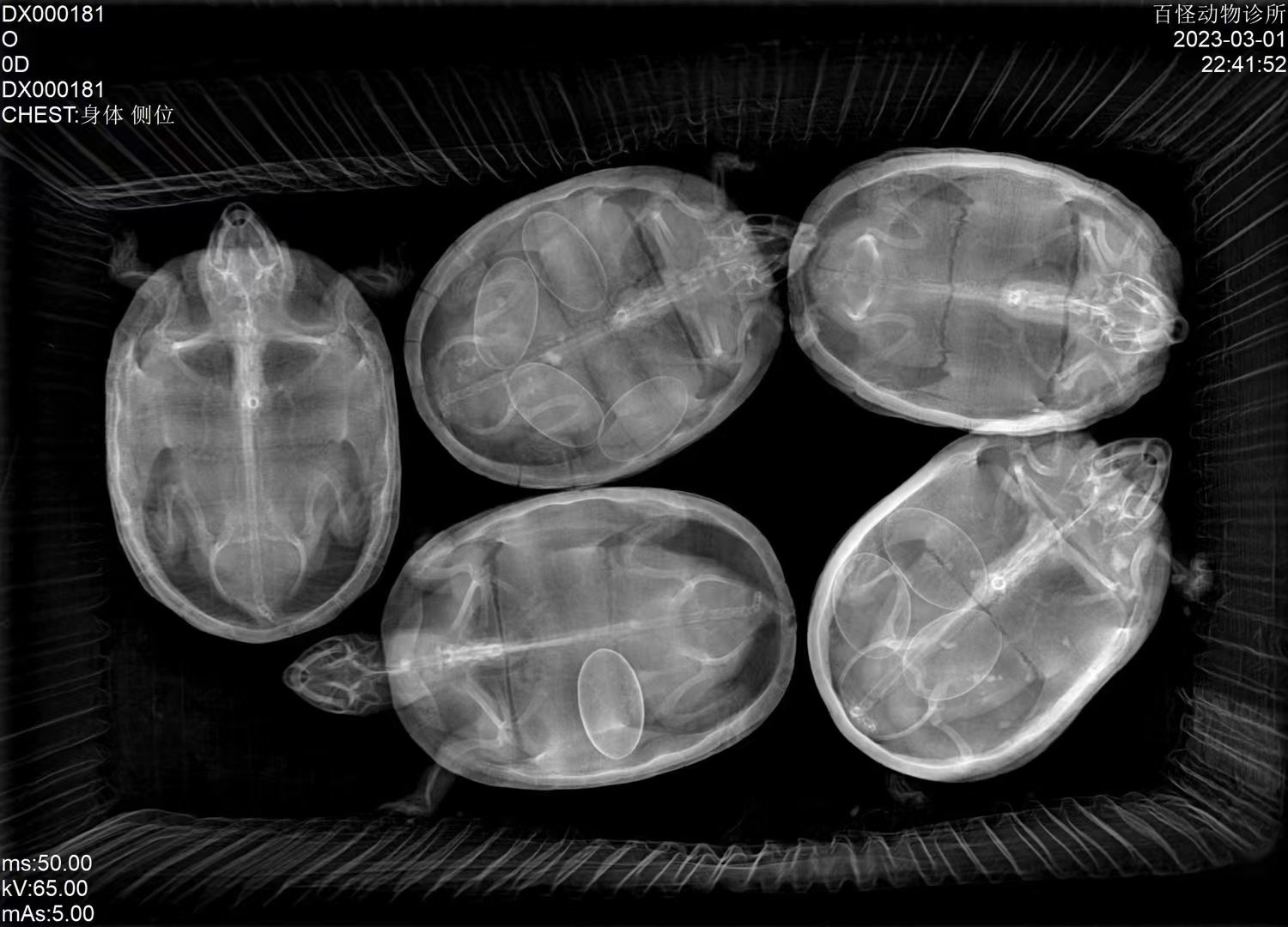
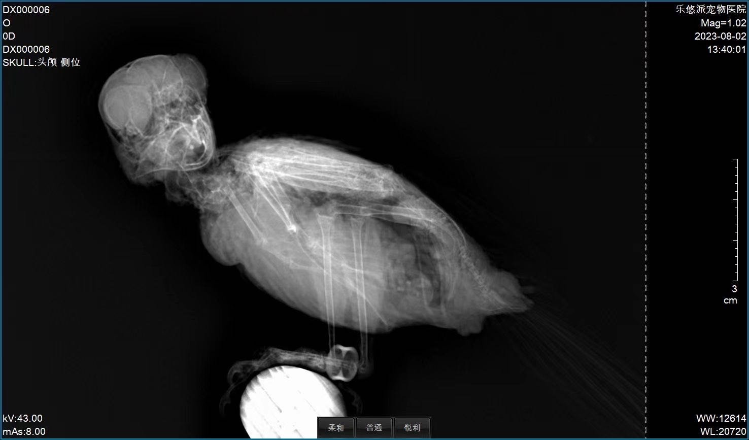
Representative radiographs across companion-animal and exotic species, illustrating image quality at the 0.6 mm fine-focus and 16-bit detector pairing.
Workflow & Installation
Single-phase 110/220 VAC supply. The removable column lets you reconfigure the room between tabletop and wall-stand workflows without re-installation. Operating environment 5–35 °C, 10–75 % RH. Engineer-led installation, calibration, and operator training included.
iPet-400 Pro removable-column installation walkthrough (English narration, ~4 min)
Package Contents
- High-voltage generator + control box
- X-ray tube assembly (Canon E7240X) + high-voltage cables
- 17×17″ CsI flat-panel detector + adapter
- LED collimator
- Removable column + four-way floating patient table (1400×700 mm)
- iVetXray workstation: 23.8″ display, 21.5″ touchscreen, PC, software dongle
- V-shape positioning set (1 large + 1 small)
- Lead protection set: gown, hat, scarf, glasses, gloves
- Foot switch, remote control, network cable
Optional: wireless flat-panel detector; extended warranty (per year); additional V-positioning and lead-protection sets.
Downloads
Why Choose IWA-DR02
- Workflow efficiency — one room, two configurations: switch between tabletop and vertical-stand without moving equipment.
- Clinical accuracy — 0.6 mm fine focus and 16-bit CsI detector preserve fine bone and dental detail.
- Practice economics — single installation covers more case types, reducing the need for a second room or system.
Warranty & Support
Two-year warranty on hardware and software. Optional extended warranty available per year. Engineer-led on-site installation, calibration, and training included.
FAQ
Q: What does the removable column enable?
You can swing the tube head off the table and rotate for vertical-stand imaging — useful for large-breed dogs, standing exams, or equine extremities.
Q: How is DR02 different from DR01?
DR02 adds the removable column, a 0.6 / 1.2 mm finer dual-focus tube (vs DR01’s 1.0 / 2.0 mm), and supports an optional wireless detector.
Q: Can the wireless detector be added later?
Yes. The optional wireless detector can be ordered with the system or retrofitted on-site.
Q: Is the workstation DICOM compliant?
Yes. iVetXray supports DICOM 3.0 Store, Print, and Worklist for major PACS systems.
Q: Does the system support large-breed dogs?
Yes. The 1400×700 mm table top and 32 kW generator handle large-breed canine and small equine extremity studies.
Reviews
There are no reviews yet.