
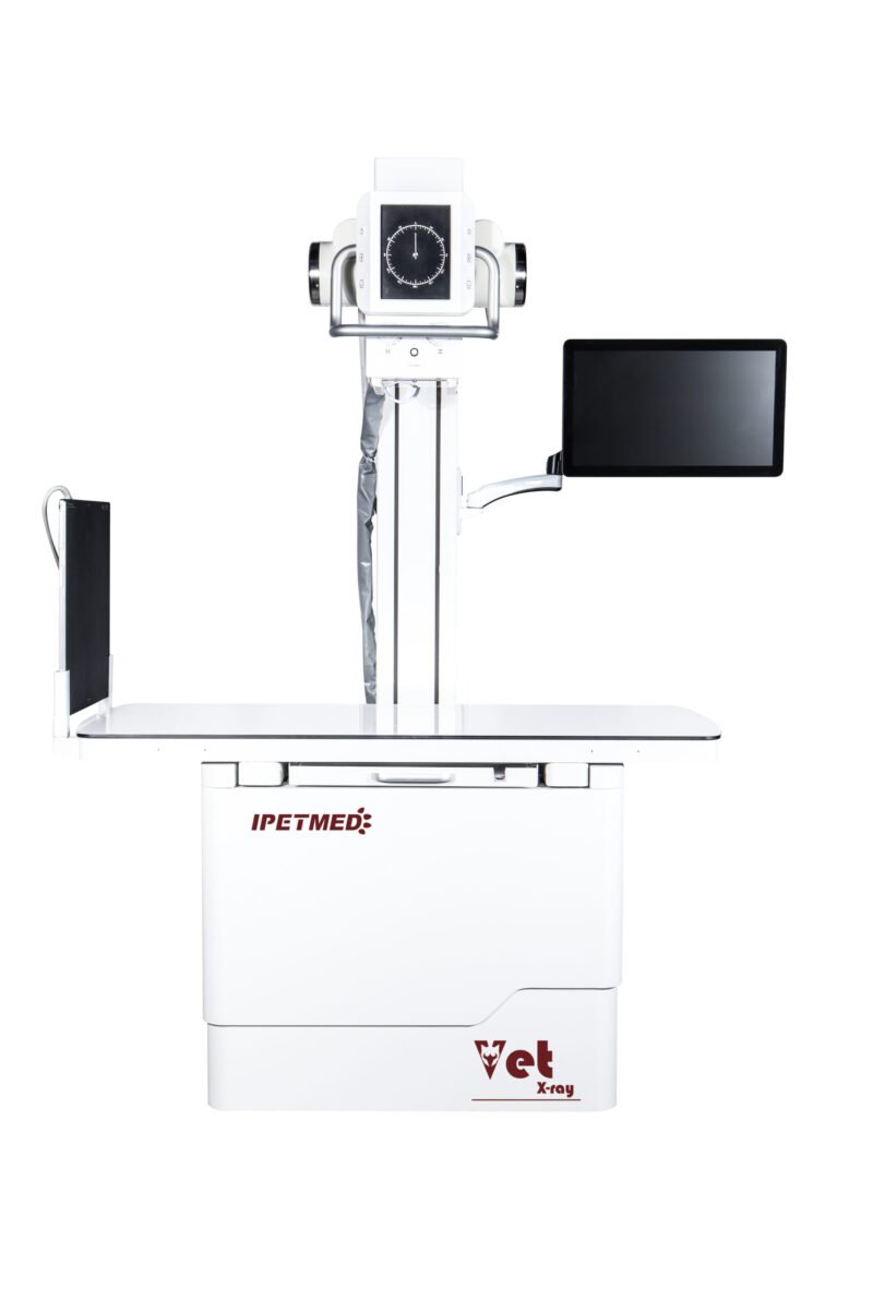
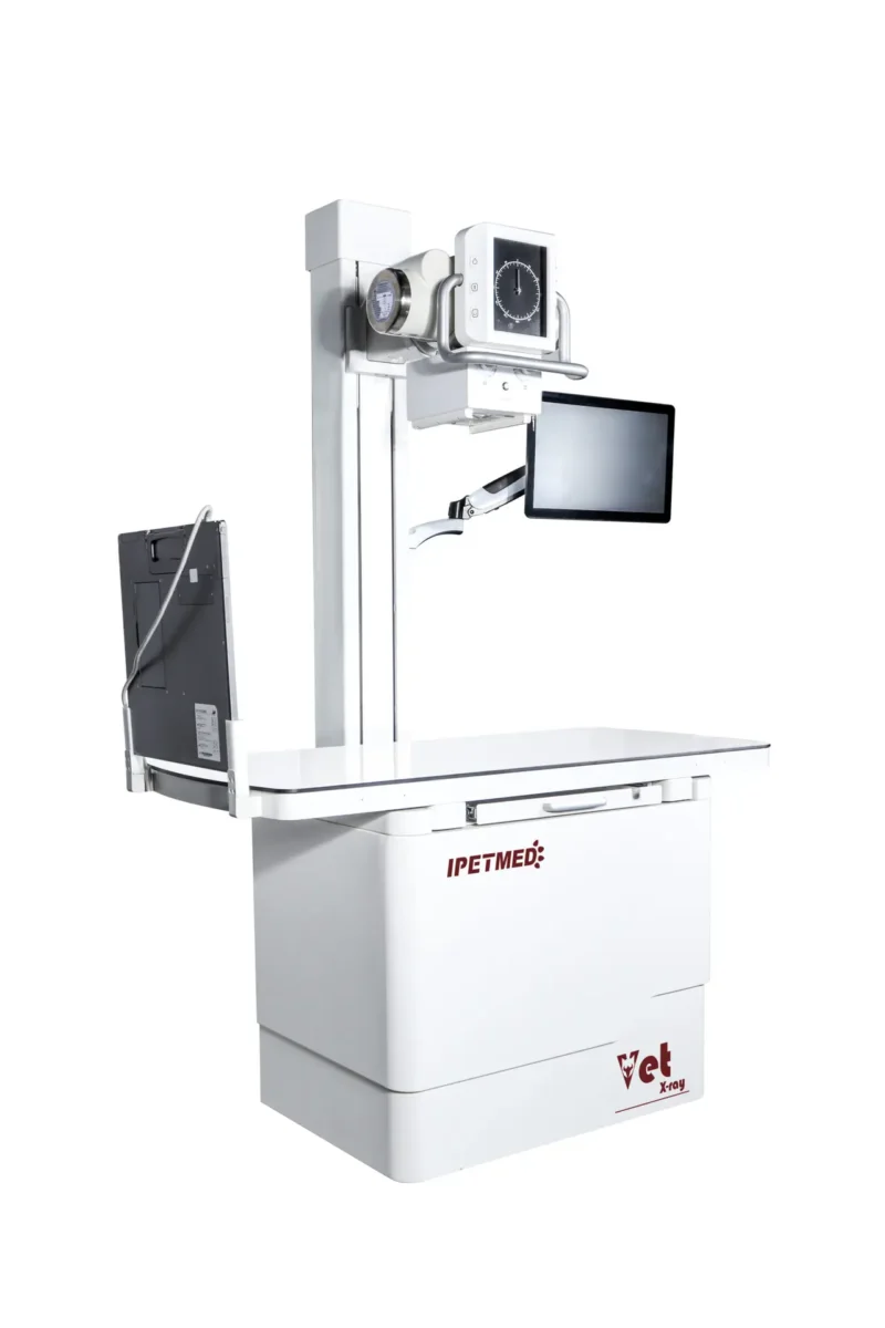
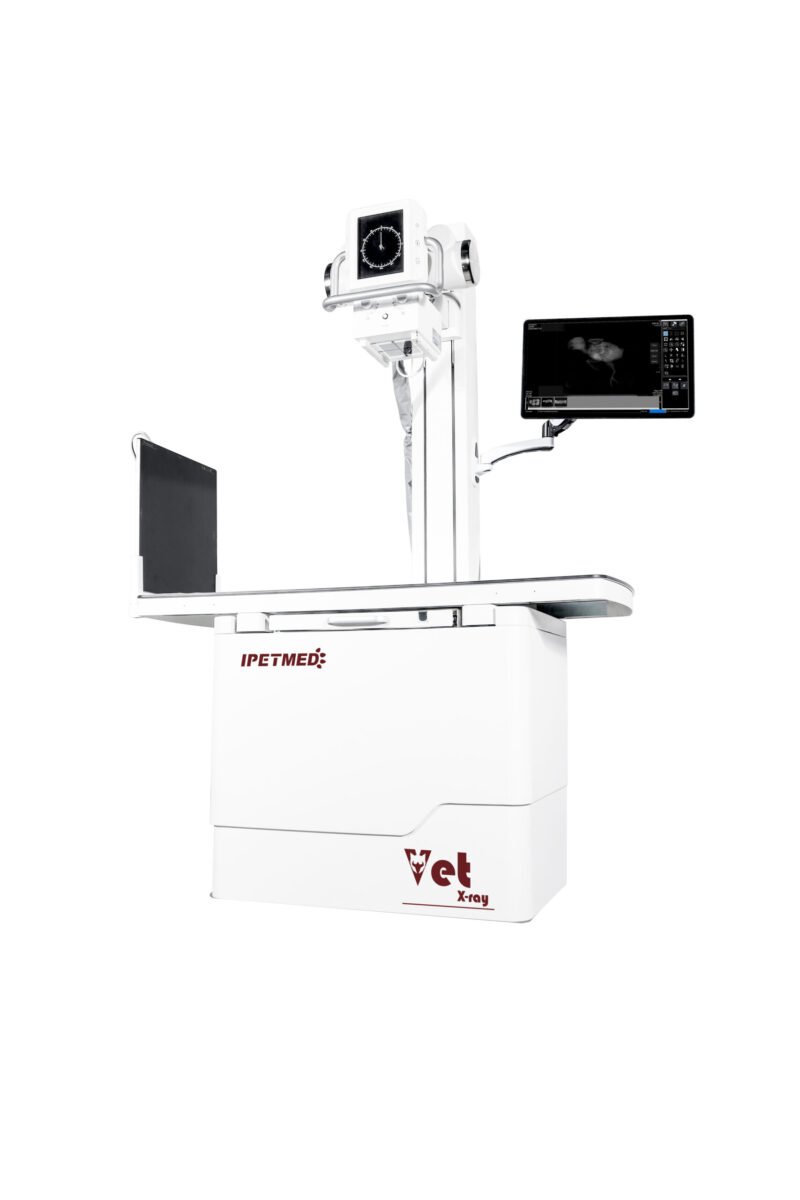
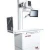
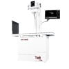
Pro-tier dynamic veterinary DR — adds a removable column for vertical-stand imaging on top of the iSmart-32 fluoroscopy platform.
Product Overview
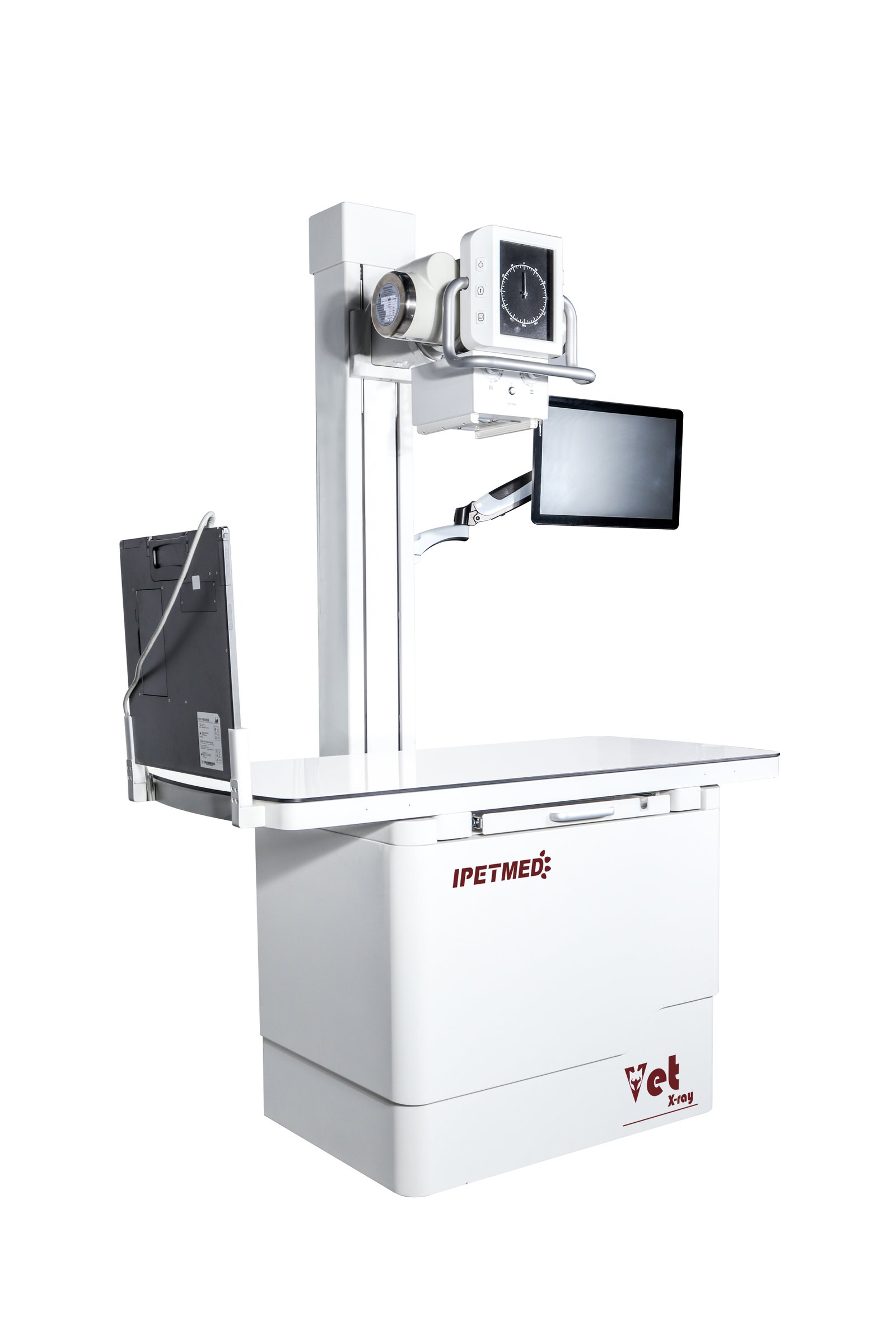
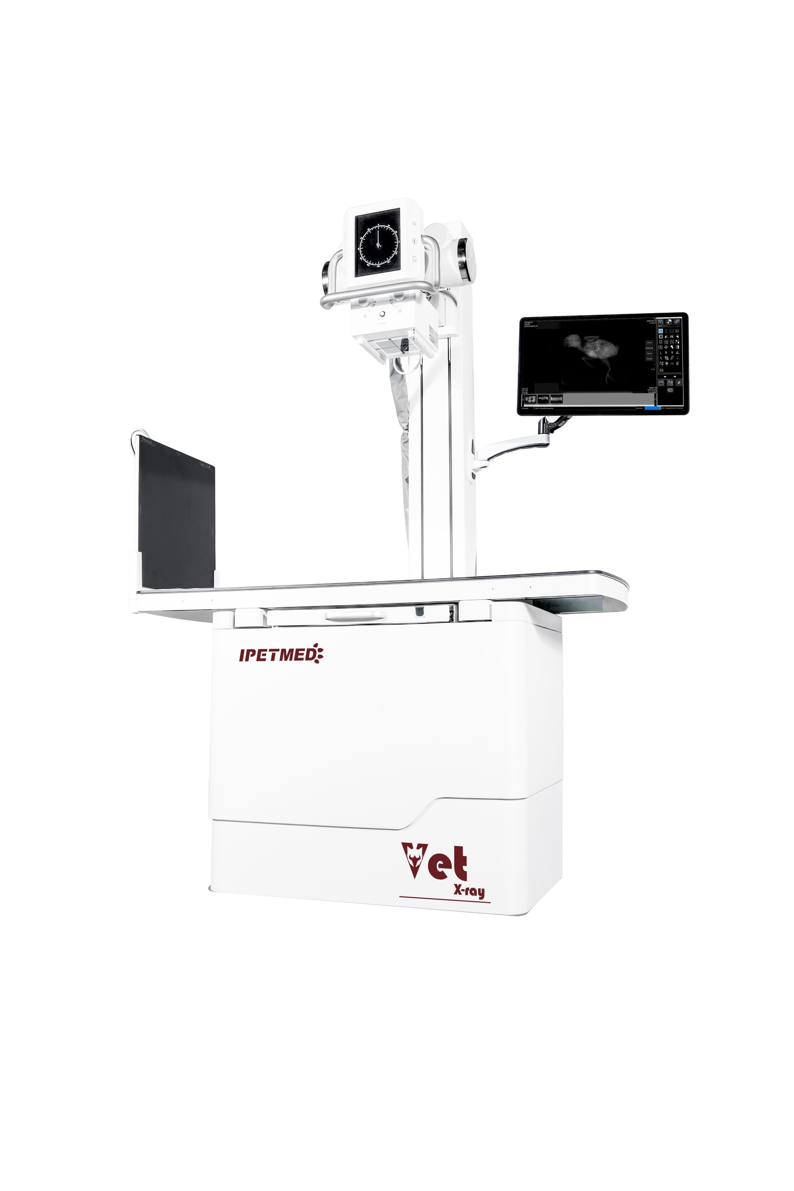
IWA-DR05 iSmart-32 Pro Multifunctional Dynamic DR System extends the iSmart-32 fluoroscopy platform with a removable column that lets the X-ray tube rotate for vertical-stand imaging — useful for standing large-breed exams, hip surveys, and equine distal-limb work. The system retains the 17×17″ dynamic CsI flat-panel detector, Canon E7240X dual-focus tube (0.6 / 1.2 mm), and 32 kW ultra-high-frequency generator that support up to 28 fps fluoroscopy alongside still radiography.
iSmart-32 Pro is positioned for hospitals that need real-time fluoroscopy plus the flexibility to handle tabletop, standing, and vertical-bucky exams in the same room without re-installation.
Key Features
- Removable column with rotating tube head — switch between tabletop radiography and vertical-stand imaging without re-installation.
- Real-time fluoroscopy at up to 28 fps with continuous and pulsed modes; DSA digital subtraction angiography included.
- Canon E7240X X-ray tube — 0.6 / 1.2 mm dual focus, 140 kHU anode heat capacity, suited to fine bone work and high-throughput exposure.
- 32 kW ultra-high-frequency generator: radiography 40–125 kV / 10–400 mA; fluoroscopy 40–110 kV continuous / pulse.
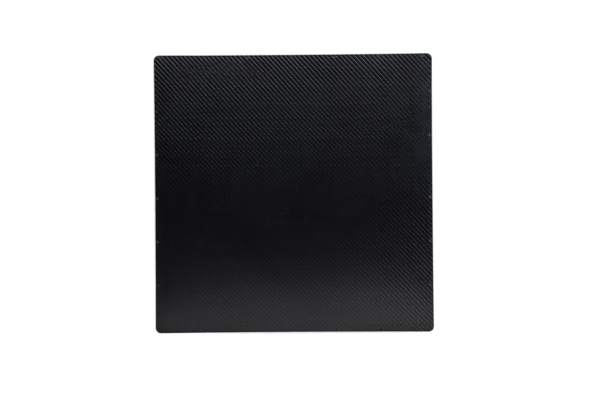
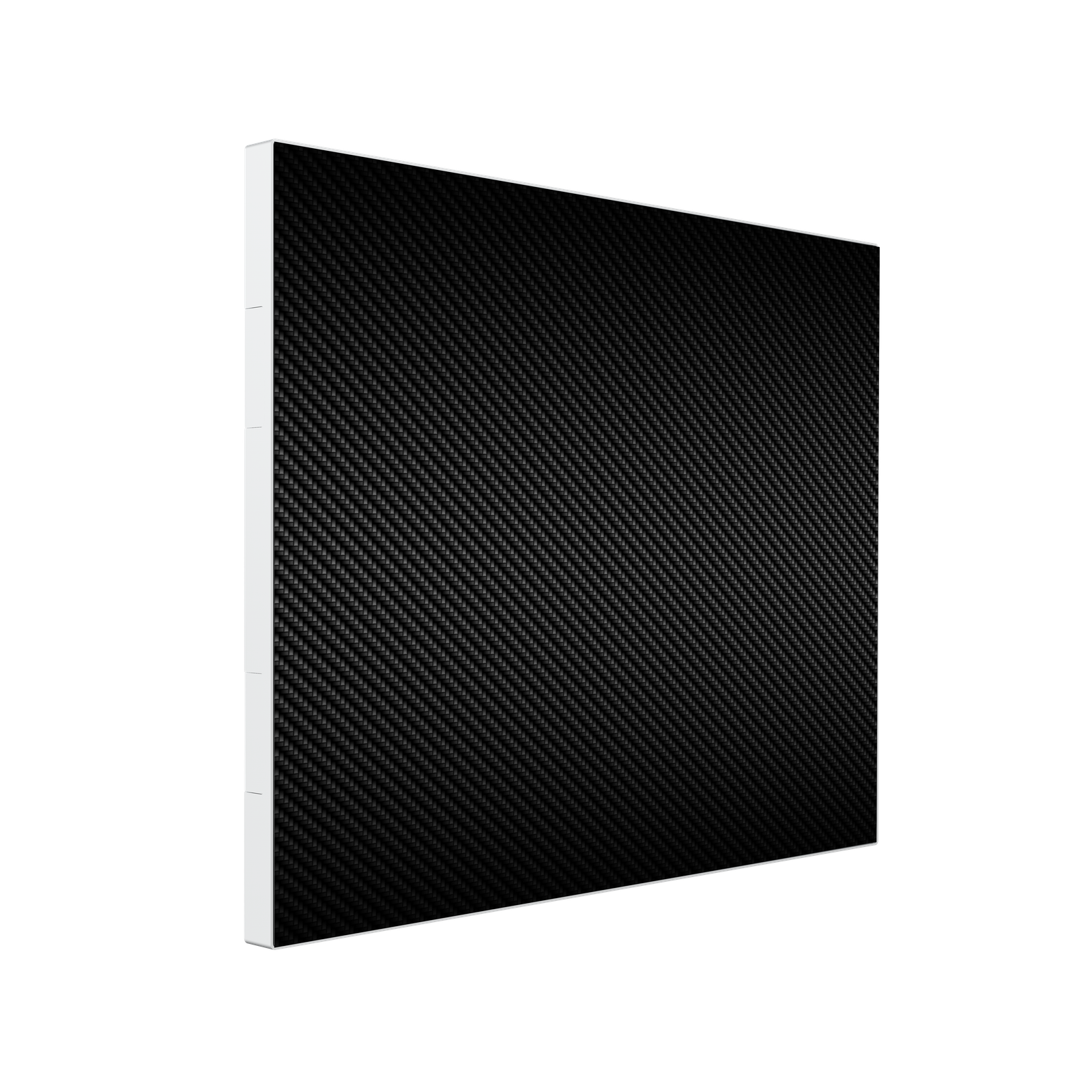
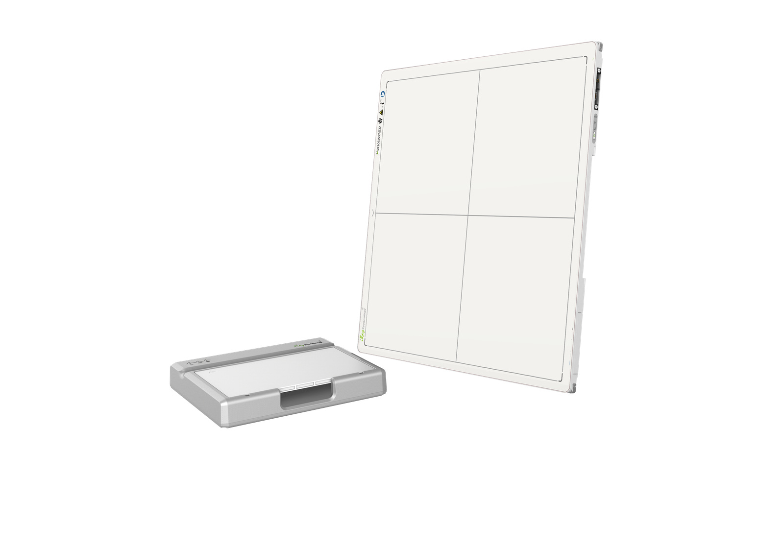
- 17×17″ dynamic CsI flat-panel detector (IWA1717DZ): 3072×3072, 139 µm, 16-bit, binning 1×1 / 2×2 / 3×3.
- Four-way floating patient table — 1400×700 mm phenolic top, X ±75 mm / Y ±200 mm travel.
- iPet Vet imaging software with size-preset APR and full veterinary measurement toolkit (VHS, TPLO, HIP, TTA, TTA2).
- Cloud imaging across multiple consultation rooms, tablets, and phones.
- 2-year warranty on hardware and software, optional extended-warranty service.
Product Gallery
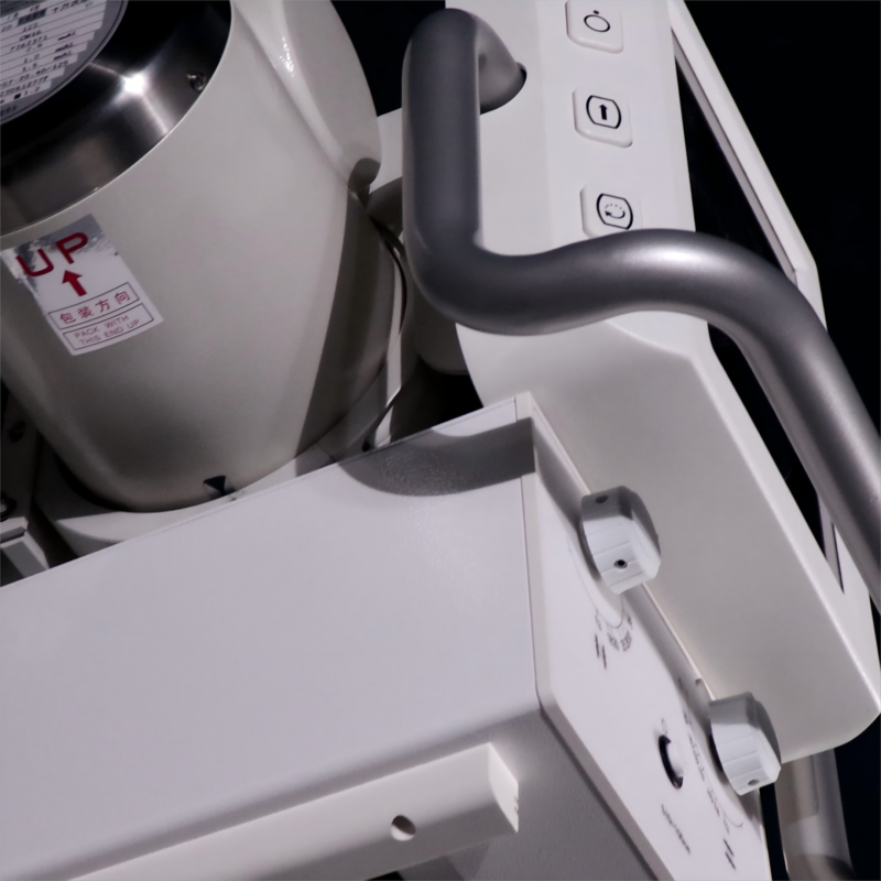
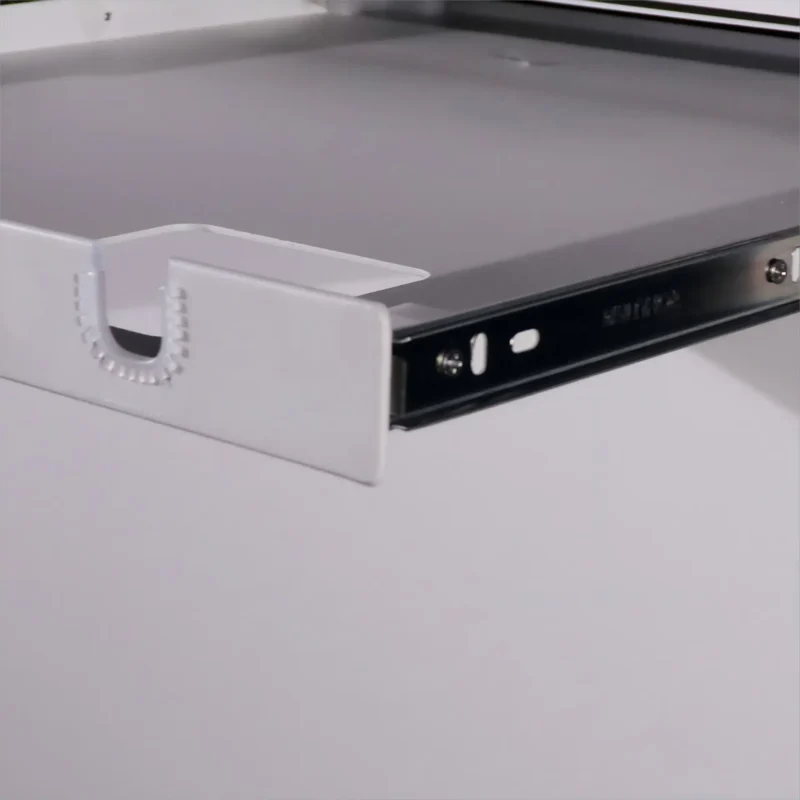
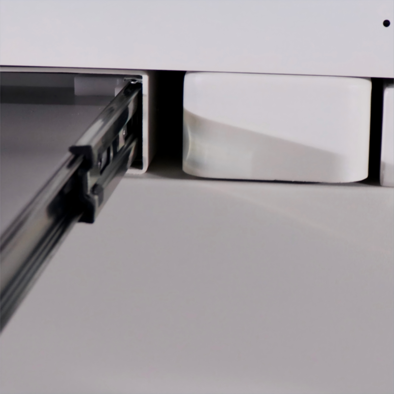
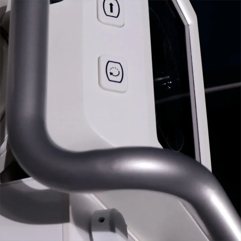
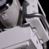
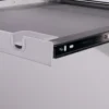
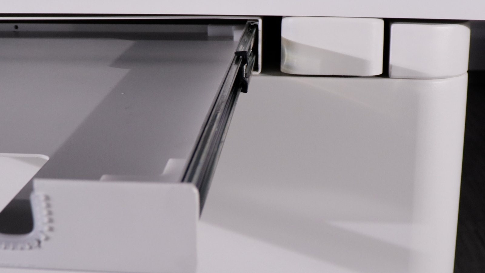
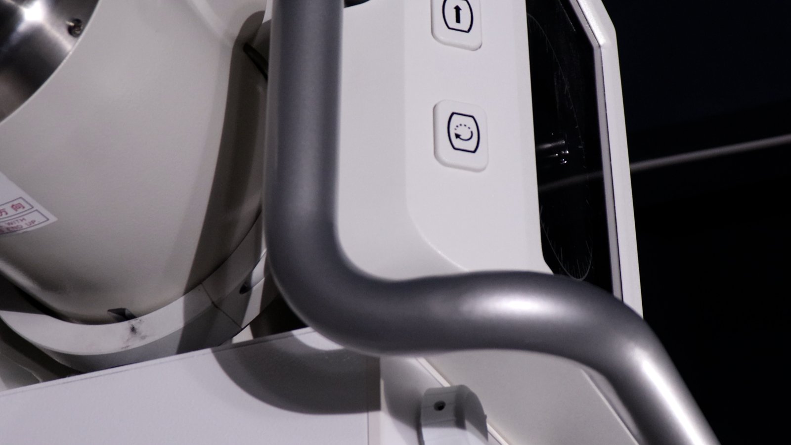
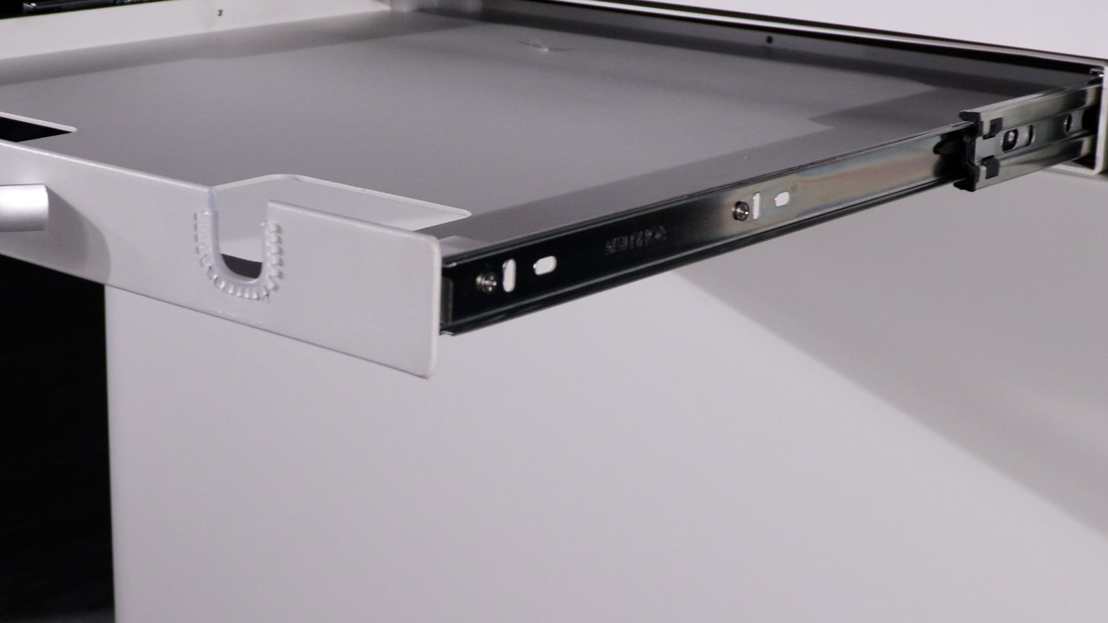
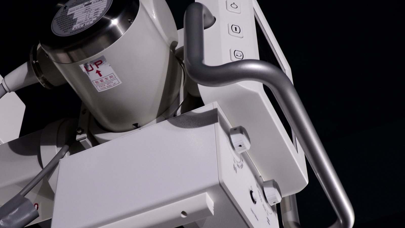
Detail views of the removable column, rotating X-ray tube head, four-way floating patient table, operator workstation, and vertical-stand imaging configuration.
High-voltage generator
| Output power | 32 kW |
| Photography kV | 40 – 125 kV |
| Fluoroscopy kV | 40 – 110 kV |
| Photography mA | 10 – 400 mA |
| Continuous fluoroscopy mA | 0.5 – 10 mA |
| Pulsed fluoroscopy mA | 0.5 – 100 mA |
| Photography exposure | 0.001 – 20 s |
| Fluoroscopy duration | up to 5 min continuous |
| Working frequency | 80 – 300 kHz |
| Power input | Single phase 220 VAC ±10%, separate 4 mm² supply line |
X-ray tube (Canon E7240X)
| Max voltage | 125 kV |
| Focal spot | 0.6 / 1.2 mm (dual) |
| Anode input power | 15 kW (small) / 30 kW (large) |
| Anode heat capacity | 140 kHU |
Dynamic flat-panel detector (IWA1717DZ)
| Sensor technology | a-Si TFT / PD matrix, CsI(Tl) needle scintillator |
| Active area | 427 × 427 mm |
| Pixel matrix | 3072 × 3072 |
| Pixel pitch | 139 µm |
| A/D conversion | 16 bit |
| Binning modes | 1×1, 2×2, 3×3 |
| Max frame rate | 4 fps (1×1), 14 fps (2×2), 28 fps (3×3) — free-run |
| Spatial resolution | 3.4 LP/mm (1×1), 1.8 LP/mm (2×2), 1.0 LP/mm (3×3) |
| Lifetime dose | ≥ 1000 Gy |
| Data interface | GigE |
Column, table, collimator
| Column | Removable; tube rotating for vertical-film imaging; SID adjustable |
| Table size | 1400 × 700 mm, phenolic resin top |
| Table movement | X: 75 ±10 mm, Y: 200 ±10 mm |
| Collimator | LED light, ABS auto-brightness in fluoroscopy mode |
| Filter | 1.0 mm Al at 75 kV |
Workstation
| Software | iVetXray (acquisition) + MiniPACS (viewer); DICOM 3.0 PACS |
| Display | 23.8″ diagnostic monitor + 21.5″ tableside touchscreen |
| OS | Windows 10 |
| Storage | 256 GB SSD + 2 TB HDD |
Clinical Applications
- Respiratory dynamics — tracheal collapse, dynamic airway evaluation in large-breed canines
- GI series — foreign body localisation, intussusception, contrast transit studies
- Orthopaedics — intra-operative positioning, hip dysplasia, TPLO / TTA, equine extremity
- Cardiovascular — cardiac silhouette evaluation, DSA angiography
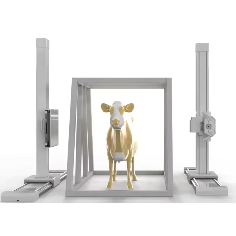
- Standing radiography of large-breed dogs or equine distal limb (with column removed)
- Static radiography — full thoracic, abdominal, skeletal, and dental imaging
Clinical Imaging Samples
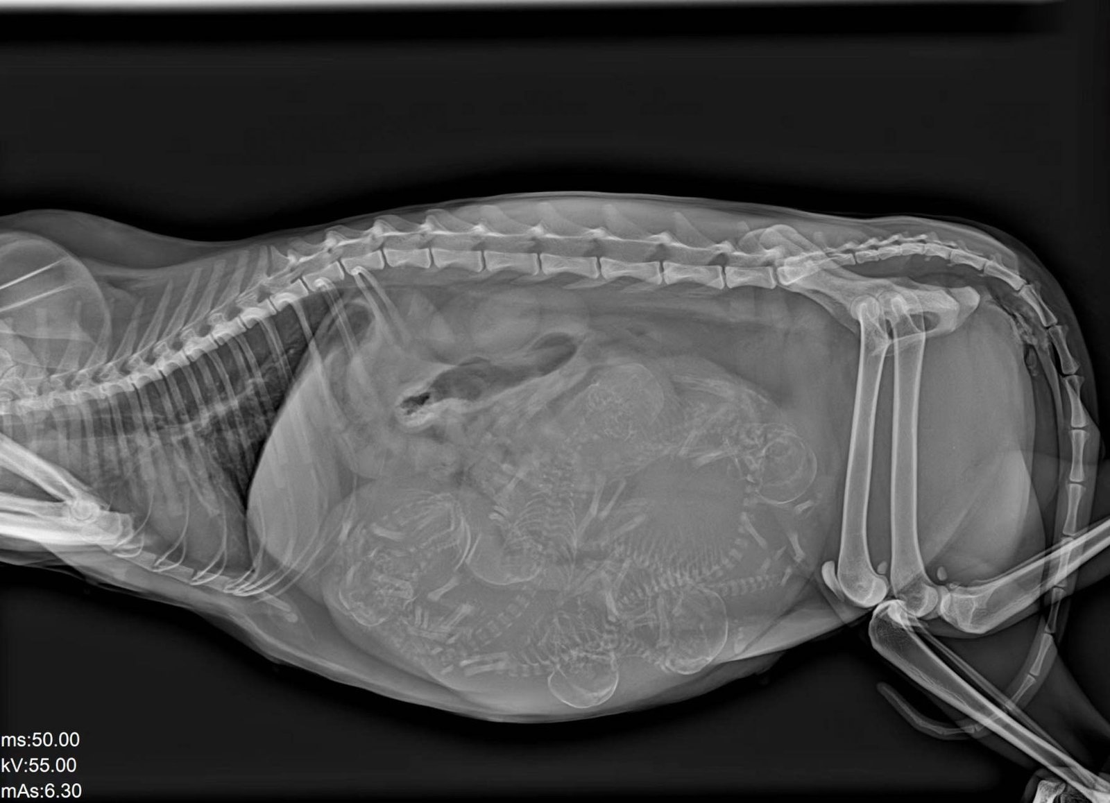
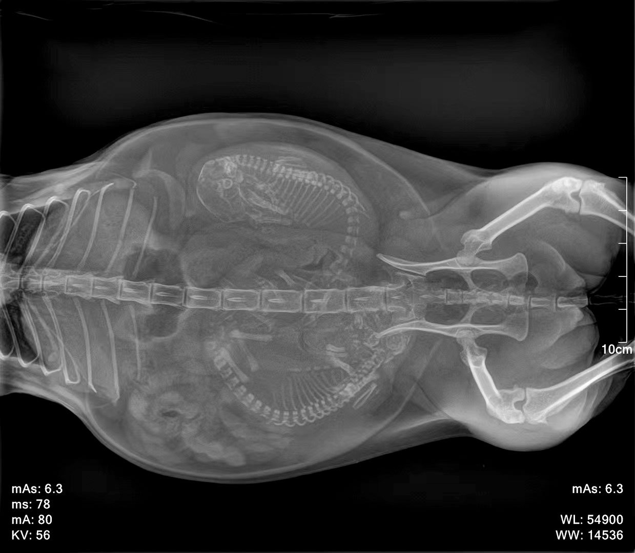
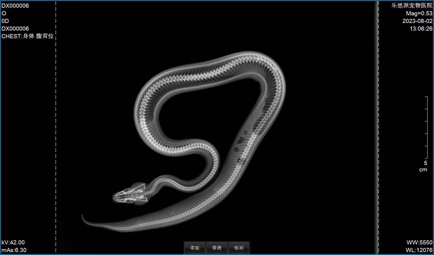
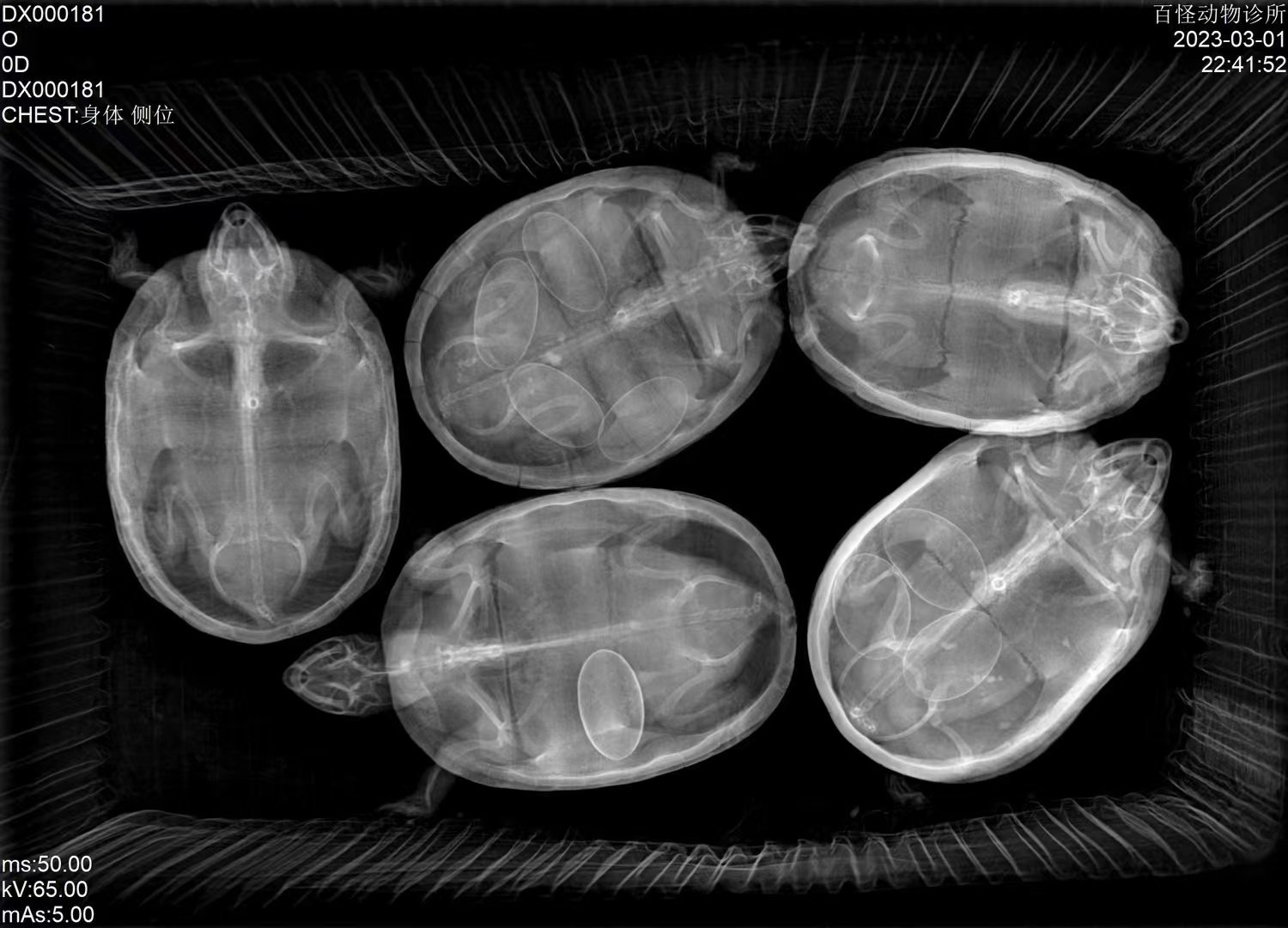
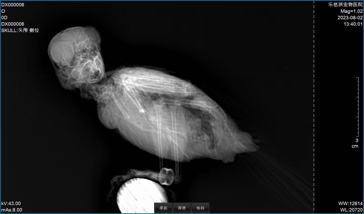
Representative still radiographs from the iSmart-32 Pro image chain across companion-animal and exotic species. Live fluoroscopy demonstration is shown below in the workflow section.
Workflow & Installation
Single-phase 220 VAC ±10 % on a dedicated 4 mm² supply line, properly grounded. The removable column lets you reconfigure the room between tabletop and vertical-stand workflows without re-installation. Operating environment 5–35 °C, 10–75 % RH. Engineer-led installation, calibration, and clinical-imaging training included.
iSmart-32 Pro live dynamic DR fluoroscopy demonstration (27 s)
Package Contents
- High-voltage generator + control box
- X-ray tube assembly (Canon E7240X) + high-voltage cables
- 17×17″ dynamic CsI flat-panel detector (IWA1717DZ) + cabling
- LED collimator with ABS auto-brightness
- Removable column + four-way floating patient table (1400×700 mm)
- iVetXray workstation: 23.8″ diagnostic display, 21.5″ tableside touchscreen, PC, software dongle
- V-shape positioning set (1 large + 1 small)
- Lead protection set: gown, hat, scarf, glasses, gloves
- Foot switch, remote control, network cable
Downloads
Why Choose IWA-DR05
- Workflow efficiency — fluoroscopy, static DR, tabletop, and vertical-stand exams in a single platform.
- Clinical accuracy — dynamic CsI detector preserves real-time motion fidelity for cardiovascular and GI workups; 0.6 mm fine focus retains detail in bone and dental work.
- Practice economics — one system covers the case mix of multiple modalities; removable column adds vertical-stand capability without a second room.
Warranty & Support
Two-year warranty on hardware and software (consumables and operator-caused damage excluded). Optional extended warranty at USD 3000 / year. 24/7 multi-engineer VIP service network. Live online clinical-imaging training courses run twice monthly through 2026.
FAQ
Q: What does iSmart-32 Pro add over iSmart-32?
A removable column with rotating tube head that lets the system handle vertical-stand imaging in addition to tabletop radiography and fluoroscopy.
Q: Can the Pro do equine extremity work?
Yes. With the column removed, the system can support standing equine distal-limb imaging — see also IWA-DR07 (iVision-1000X) for full-body equine imaging.
Q: Is DSA included as standard?
Yes. Digital subtraction angiography is a standard software feature of the dynamic detector.
Q: How long can continuous fluoroscopy run?
Up to 5 minutes continuous; pulsed mode for longer interventional procedures.
Q: Does the system require a dedicated three-phase supply?
No. iSmart-32 Pro runs on single-phase 220 VAC ±10 % via a dedicated 4 mm² supply line.
Reviews
There are no reviews yet.